
Page 1279 - Small Animal Internal Medicine, 6th Edition
P. 1279
CHAPTER 73 Common Immune-Mediated Diseases 1251
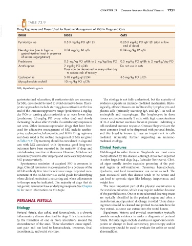
TABLE 73.9
VetBooks.ir Drug Regimens and Doses Used for Routine Management of MG in Dogs and Cats
DRUG
CATS
DOGS
Pyridostigmine 0.5-3 mg/kg PO q8-12h 0.25-3 mg/kg PO q8-12h (start at low
end of dose)
Neostigmine (use to bypass 0.04 mg/kg IM q6h 0.04 mg/kg IM q6h
gastrointestinal tract in presence
of severe regurgitation)
Prednisone 0.5 mg/kg PO q48h to 2 mg/kg/day PO 0.5 mg/kg PO q48h to 2 mg/kg/day PO
Azathioprine 2 mg/kg PO q24h Do not use in cats
Dose can be decreased to every other day
to reduce risk of toxicity
Cyclosporine 5-10 mg/kg q12-24h 3-5 mg/kg PO q12h
Mycophenolate mofetil 10 mg/kg PO q12h
MG, Myasthenia gravis.
gastrointestinal ulceration; if corticosteroids are necessary The etiology is not fully understood, but the majority of
for MG, care should be used to avoid excessive doses. Thera- evidence supports an immune-mediated mechanism. Histo-
peutic approaches include starting glucocorticoids at the low logically, affected tissues are infiltrated by lymphocytes and
end of the immunosuppressive range (prednisone 2 mg/kg/ plasma cells (primarily secreting IgA and IgG), as well as
day PO) or starting glucocorticoids at an even lower dose eosinophils and macrophages. The lymphocytes in these
(prednisone 0.5 mg/kg PO every other day) and slowly tissues are predominantly T cells, with high concentrations
increasing the dose after 2 weeks if a satisfactory response is of IL-2 and tumor necrosis factor-α present, indicating a
not seen. Other immunosuppressive drugs that have been cell-mediated immune response. German Shepherds are the
used for adjunctive management of MG include azathio- most common breed to be diagnosed with perianal fistulas,
prine, cyclosporine, leflunomide, and MMF. Drug regimens and this breed is known to have an impairment in cell-
and doses used in the routine management of MG are given mediated immunity, further supporting an immune-
in Table 73.9. Thymectomy should be considered in dogs and mediated etiology.
cats with MG associated with thymoma; good long-term
outcomes have been reported in the majority of dogs and Clinical Features
cats following resection of thymoma. However, MG does not Middle-aged to older German Shepherds are most com-
consistently resolve after surgery, and some cats may develop monly affected by this disease, although it has been reported
MG postoperatively. in other large-breed dogs (e.g., Labrador Retrievers). Clini-
Spontaneous remission of acquired MG is common in cal signs usually involve excessive grooming of the peri-
dogs. Clinical remission is accompanied by a decrease of the anal region or self-mutilation. Tenesmus, hematochezia,
AChR antibody titer into the reference range. Repeated mea- dyschezia, and fecal incontinence can occur as well. The
surement of the AChR titer is a useful guide for identifying pain associated with this disease tends to be severe and
when clinical remission is occurring and when adjustments can lead to systemic signs like lethargy, inappetance, and
to therapy may be indicated. The majority of dogs that do weight loss.
not go into remission have underlying neoplasia. See Chapter The most important part of the physical examination is
66 for more information on this topic. the rectal examination, which may require sedation because
of the painful lesions. One or more ulcerated draining tracts
are typically identified in the perianal region and often a
PERIANAL FISTULA malodorous, mucopurulent discharge is noted. These drain-
ing tracts should be cleaned and probed to evaluate how far
Etiology they extend, as some can extend into the rectal lumen.
Perianal fistula, also called anal furunculosis, is a chronic Signalment, history, and physical examination typically
inflammatory disease described in dogs. It is characterized provide enough evidence to make a diagnosis of perianal
by the formation of one or more ulcerations around the fistula. If clinical signs include diarrhea or significant consti-
circumference of the anus. These ulcerations cause signifi- pation or change in fecal consistency, proctoscopy and/or
cant pain and can lead to hematochezia, tenesmus, fecal colonoscopy should be used to evaluate for colitis or rectal
incontinence, and rectal stricture. abnormalities.