
Page 58 - Small Animal Internal Medicine, 6th Edition
P. 58
30 PART I Cardiovascular System Disorders
pulmonary venous flow patterns, and others (see Suggested of blood flow velocity and direction occurs all along the
Readings for more information). ultrasound beam, not in a specified area (so-called range
VetBooks.ir accelerates rapidly during ejection with more gradual decel- ambiguity).
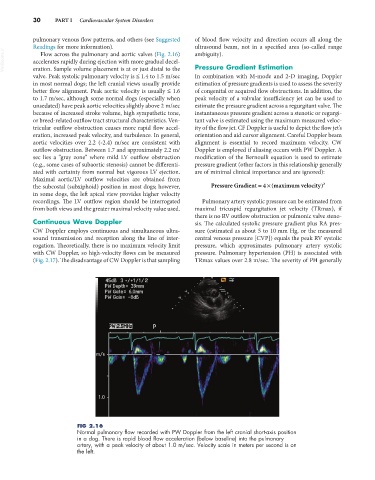
Flow across the pulmonary and aortic valves (Fig. 2.16)
Pressure Gradient Estimation
eration. Sample volume placement is at or just distal to the
valve. Peak systolic pulmonary velocity is ≤ 1.4 to 1.5 m/sec In combination with M-mode and 2-D imaging, Doppler
in most normal dogs; the left cranial views usually provide estimation of pressure gradients is used to assess the severity
better flow alignment. Peak aortic velocity is usually ≤ 1.6 of congenital or acquired flow obstructions. In addition, the
to 1.7 m/sec, although some normal dogs (especially when peak velocity of a valvular insufficiency jet can be used to
unsedated) have peak aortic velocities slightly above 2 m/sec estimate the pressure gradient across a regurgitant valve. The
because of increased stroke volume, high sympathetic tone, instantaneous pressure gradient across a stenotic or regurgi-
or breed-related outflow tract structural characteristics. Ven- tant valve is estimated using the maximum measured veloc-
tricular outflow obstruction causes more rapid flow accel- ity of the flow jet. CF Doppler is useful to depict the flow jet’s
eration, increased peak velocity, and turbulence. In general, orientation and aid cursor alignment. Careful Doppler beam
aortic velocities over 2.2 (-2.4) m/sec are consistent with alignment is essential to record maximum velocity. CW
outflow obstruction. Between 1.7 and approximately 2.2 m/ Doppler is employed if aliasing occurs with PW Doppler. A
sec lies a “gray zone” where mild LV outflow obstruction modification of the Bernoulli equation is used to estimate
(e.g., some cases of subaortic stenosis) cannot be differenti- pressure gradient (other factors in this relationship generally
ated with certainty from normal but vigorous LV ejection. are of minimal clinical importance and are ignored):
Maximal aortic/LV outflow velocities are obtained from
4
the subcostal (subxiphoid) position in most dogs; however, Pressure Gradient =×( maximum velocity) 2
in some dogs, the left apical view provides higher velocity
recordings. The LV outflow region should be interrogated Pulmonary artery systolic pressure can be estimated from
from both views and the greater maximal velocity value used. maximal tricuspid regurgitation jet velocity (TRmax), if
there is no RV outflow obstruction or pulmonic valve steno-
Continuous Wave Doppler sis. The calculated systolic pressure gradient plus RA pres-
CW Doppler employs continuous and simultaneous ultra- sure (estimated as about 5 to 10 mm Hg, or the measured
sound transmission and reception along the line of inter- central venous pressure [CVP]) equals the peak RV systolic
rogation. Theoretically, there is no maximum velocity limit pressure, which approximates pulmonary artery systolic
with CW Doppler, so high-velocity flows can be measured pressure. Pulmonary hypertension (PH) is associated with
(Fig. 2.17). The disadvantage of CW Doppler is that sampling TRmax values over 2.8 m/sec. The severity of PH generally
FIG 2.16
Normal pulmonary flow recorded with PW Doppler from the left cranial short-axis position
in a dog. There is rapid blood flow acceleration (below baseline) into the pulmonary
artery, with a peak velocity of about 1.0 m/sec. Velocity scale in meters per second is on
the left.