
Page 859 - Small Animal Internal Medicine, 6th Edition
P. 859
CHAPTER 49 Disorders of the Endocrine Pancreas 831
VetBooks.ir
A
B C
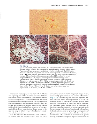
FIG 49.12
(A) Severe islet amyloidosis (black arrow) in a cat with initial non–insulin-dependent
diabetes mellitus (NIDDM) that progressed to insulin-dependent diabetes mellitus (IDDM).
A pancreatic biopsy specimen was obtained while the animal was in the IDDM state.
Residual β cells containing insulin (red arrows) are also present. (Immunoperoxidase stain,
×100.) (B) Severe vacuolar degeneration of islet cells. Pancreatic tissue was evaluated at
necropsy 28 months after diabetes was diagnosed and 20 months after the cat
progressed from NIDDM to IDDM, requiring insulin to control blood glucose
concentrations. The cat died from metastatic exocrine pancreatic adenocarcinoma.
(Hematoxylin and eosin stain; ×500.) (C) Severe chronic pancreatitis with fibrosis in a
diabetic cat with IDDM. The cat was euthanized because of persistent problems with
lethargy, inappetence, and poorly controlled diabetes mellitus. (Hematoxylin and eosin
stain; ×100.) (A from Feldman EC et al: Canine and feline endocrinology and
reproduction, ed 3, St Louis, 2004, WB Saunders.)
Glucose toxicity also plays an important role in diabetic exposed to a concurrent insulin-antagonistic drug or disease,
cats, especially as it relates to diabetic remission. Diabetic most notably glucocorticoids and chronic inflammation
remission is defined as a scenario in which diabetes mellitus (Fig. 49.13). Unlike healthy cats, those that experience dia-
is correctly diagnosed in a cat, insulin treatment is initiated betic remission have a reduced population of β cells, dys-
in conjunction with adjustments in diet and discontinuation functional β cells, or both, and this impairs the ability of the
of insulin antagonistic medications (most notably glucocor- pancreas to compensate for concurrent insulin resistance.
ticoids) and, weeks to months later, hyperglycemia resolves An inadequate insulin response results in hyperglycemia.
and insulin is discontinued without recurrence of hypergly- Persistent hyperglycemia can, in turn, cause hypoinsulinemia
cemia. Blood glucose concentrations should remain in the by suppressing the function of remaining β cells and can
reference range for at least 4 weeks before diabetes is consid- induce insulin resistance by promoting downregulation of
ered to be in remission. glucose transport systems, causing a defect in posttransport
Cats that experience diabetic remission are in a subclini- insulin action. This phenomenon is referred to as glucose
cal diabetic state that becomes clinical when the cat is toxicity. β cells have an impaired response to stimulation by