
Page 870 - Small Animal Internal Medicine, 6th Edition
P. 870
842 PART VI Endocrine Disorders
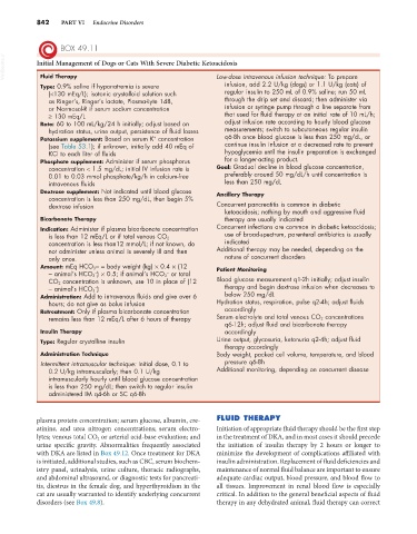
BOX 49.11
VetBooks.ir Initial Management of Dogs or Cats With Severe Diabetic Ketoacidosis
Fluid Therapy
Low-dose intravenous infusion technique: To prepare
Type: 0.9% saline if hyponatremia is severe infusion, add 2.2 U/kg (dogs) or 1.1 U/kg (cats) of
(<130 mEq/L); isotonic crystalloid solution such regular insulin to 250 mL of 0.9% saline; run 50 mL
as Ringer’s, Ringer’s lactate, Plasma-Lyte 148, through the drip set and discard; then administer via
or Normosol-R if serum sodium concentration infusion or syringe pump through a line separate from
≥ 130 mEq/L that used for fluid therapy at an initial rate of 10 mL/h;
Rate: 60 to 100 mL/kg/24 h initially; adjust based on adjust infusion rate according to hourly blood glucose
hydration status, urine output, persistence of fluid losses measurements; switch to subcutaneous regular insulin
+
Potassium supplement: Based on serum K concentration q6-8h once blood glucose is less than 250 mg/dL, or
(see Table 53.1); if unknown, initially add 40 mEq of continue insulin infusion at a decreased rate to prevent
KCl to each liter of fluids hypoglycemia until the insulin preparation is exchanged
Phosphate supplement: Administer if serum phosphorus for a longer-acting product.
concentration < 1.5 mg/dL; initial IV infusion rate is Goal: Gradual decline in blood glucose concentration,
0.01 to 0.03 mmol phosphate/kg/h in calcium-free preferably around 50 mg/dL/h until concentration is
intravenous fluids less than 250 mg/dL
Dextrose supplement: Not indicated until blood glucose Ancillary Therapy
concentration is less than 250 mg/dL, then begin 5%
dextrose infusion Concurrent pancreatitis is common in diabetic
ketoacidosis; nothing by mouth and aggressive fluid
Bicarbonate Therapy therapy are usually indicated
Indication: Administer if plasma bicarbonate concentration Concurrent infections are common in diabetic ketoacidosis;
is less than 12 mEq/L or if total venous CO 2 use of broad-spectrum, parenteral antibiotics is usually
concentration is less than12 mmol/L; if not known, do indicated
not administer unless animal is severely ill and then Additional therapy may be needed, depending on the
only once. nature of concurrent disorders
Amount: mEq HCO 3 − = body weight (kg) × 0.4 × (12 Patient Monitoring
−
− animal’s HCO 3 ) × 0.5; if animal’s HCO 3 or total
−
CO 2 concentration is unknown, use 10 in place of (12 Blood glucose measurement q1-2h initially; adjust insulin
−
− animal’s HCO 3 ) therapy and begin dextrose infusion when decreases to
Administration: Add to intravenous fluids and give over 6 below 250 mg/dL
hours; do not give as bolus infusion Hydration status, respiration, pulse q2-4h; adjust fluids
Retreatment: Only if plasma bicarbonate concentration accordingly
remains less than 12 mEq/L after 6 hours of therapy Serum electrolyte and total venous CO 2 concentrations
q6-12h; adjust fluid and bicarbonate therapy
Insulin Therapy accordingly
Type: Regular crystalline insulin Urine output, glycosuria, ketonuria q2-4h; adjust fluid
therapy accordingly
Administration Technique Body weight, packed cell volume, temperature, and blood
Intermittent intramuscular technique: Initial dose, 0.1 to pressure q6-8h
0.2 U/kg intramuscularly; then 0.1 U/kg Additional monitoring, depending on concurrent disease
intramuscularly hourly until blood glucose concentration
is less than 250 mg/dL; then switch to regular insulin
administered IM q4-6h or SC q6-8h
plasma protein concentration; serum glucose, albumin, cre- FLUID THERAPY
atinine, and urea nitrogen concentrations; serum electro- Initiation of appropriate fluid therapy should be the first step
lytes; venous total CO 2 or arterial acid-base evaluation; and in the treatment of DKA, and in most cases it should precede
urine specific gravity. Abnormalities frequently associated the initiation of insulin therapy by 2 hours or longer to
with DKA are listed in Box 49.12. Once treatment for DKA minimize the development of complications affiliated with
is initiated, additional studies, such as CBC, serum biochem- insulin administration. Replacement of fluid deficiencies and
istry panel, urinalysis, urine culture, thoracic radiographs, maintenance of normal fluid balance are important to ensure
and abdominal ultrasound, or diagnostic tests for pancreati- adequate cardiac output, blood pressure, and blood flow to
tis, diestrus in the female dog, and hyperthyroidism in the all tissues. Improvement in renal blood flow is especially
cat are usually warranted to identify underlying concurrent critical. In addition to the general beneficial aspects of fluid
disorders (see Box 49.8). therapy in any dehydrated animal, fluid therapy can correct