
Page 872 - Small Animal Internal Medicine, 6th Edition
P. 872
844 PART VI Endocrine Disorders
ECF ECF
ICF H H ICF H H ↑↑ ECF ICF H H ↓↓
VetBooks.ir K K K K ↑↑ K K ↓
PO 4 2 PO 4 2 PO 4 2 PO 4 2 ↑ PO 4 2 PO 4 2 ↓
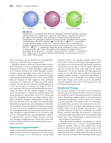
A Normal pH B Acidosis C Correction of acidosis
FIG 49.18
Redistribution of extracellular fluid (ECF) and intracellular fluid (ICF) hydrogen, potassium,
and phosphate ions in response to a decrease in ECF pH (i.e., acidosis), an increase in
ECF glucose and osmolality, and translocation of water from the ICF to the ECF
compartment and subsequent correction of acidosis and the intracellular shift of glucose
+
and electrolytes with insulin treatment. (A) Normal ECF pH. (B) ECF H concentration
+
increases during acidosis, causing H to move into cells and down its concentration
gradient. Increases in ECF glucose and osmolality cause extracellular shifts of water, K ,
+
and PO 4 . (C) ECF H concentration decreases during correction of acidosis, causing H
+
+
+2
to move out of the cells. Insulin administration and correction of acidemia cause an
+
+2
+
+2
intracellular shift of glucose, K , and PO 4 , decreasing ECF K and PO 4 concentrations.
(From Feldman EC et al: Canine and feline endocrinology and reproduction, ed 3, St
Louis, 2004, WB Saunders.)
ataxia, and seizures may be observed. Severe hypophospha- treatment of DKA, and typically normalize without treat-
temia may be clinically silent in many animals. ment as DKA resolves. Clinical signs of hypomagnesemia do
Phosphate therapy is indicated if clinical signs or hemo- not usually occur until serum total and ionized magnesium
lysis is identified or if the serum phosphorus concentration concentrations are less than 1.0 and 0.4 mg/dL, respectively,
decreases to less than 1.5 mg/dL. Phosphate is supplemented and even at these low levels many dogs and cats remain
by IV infusion. Potassium and sodium phosphate solutions asymptomatic. We do not routinely treat hypomagnesemia
contain 3 mmol of phosphate and 4.4 mEq of potassium or in dogs or cats with DKA unless problems with persistent
4 mEq of sodium per milliliter. The recommended dosage lethargy, anorexia, weakness, or refractory hypokalemia or
for phosphate supplementation is 0.01 to 0.03 mmol of phos- hypocalcemia are encountered after 24 to 48 hours of fluid
phate per kilogram of body weight per hour, preferably and insulin therapy, and another cause for the problem
administered in calcium-free IV fluids (e.g., 0.9% sodium cannot be identified. See Chapter 53 for information on the
chloride). In dogs and cats with severe hypophosphatemia, treatment of hypomagnesemia.
it may be necessary to increase the dosage to 0.03 to
0.12 mmol/kg/h. Because the dose of phosphate necessary to Bicarbonate Therapy
replete an animal and the animal’s response to therapy The clinical presentation of the dog or cat, in conjunction
cannot be predicted, it is important to initially monitor the with the plasma bicarbonate or total venous CO 2 concentra-
serum phosphorus concentration every 8 to 12 hours and tion, should be used to determine the need for bicarbonate
adjust the phosphate infusion accordingly. Adverse effects therapy. Bicarbonate supplementation is not recommended
from overzealous phosphate administration include iatro- when plasma bicarbonate (or total venous CO 2 ) is 12 mEq/L
genic hypocalcemia and its associated neuromuscular signs, or greater, especially if the animal is alert. An alert dog or
hypernatremia, hypotension, and calcification of soft tissues. cat probably has a normal or near-normal pH in the cere-
Serum ionized calcium concentration should be measured at brospinal fluid (CSF). Acidosis in these animals is corrected
the same time as serum phosphorus concentration and the through insulin and fluid therapy. Improvement in renal
rate of phosphate infusion decreased if hypocalcemia is iden- perfusion enhances urinary loss of ketoacids, and insulin
tified. Phosphorus supplementation is not indicated in dogs therapy markedly diminishes the production of ketoacids.
and cats with hypercalcemia, hyperphosphatemia, oliguria, Acetoacetate and β-hydroxybutyrate are metabolically usable
or suspected tissue necrosis. If kidney function is in ques- anions, and 1 mEq of bicarbonate is generated from each
tion, phosphorus supplementation should not be done until 1 mEq of ketoacid metabolized.
the status of kidney function and serum phosphorus concen- When the plasma bicarbonate concentration is 11 mEq/L
tration are known. or less (total venous CO 2 < 12 mEq/L), bicarbonate therapy
should be initiated. Many of these animals have severe
Magnesium Supplementation depression that may be a result of concurrent severe central
Plasma total and ionized magnesium concentrations may be nervous system acidosis. Metabolic acidosis should be cor-
within or below the reference range at the time DKA is diag- rected slowly, thereby avoiding major alterations in the pH
nosed in the dog or cat, often decrease during initial of the CSF. Only a portion of the bicarbonate deficit is given