
Page 877 - Small Animal Internal Medicine, 6th Edition
P. 877
CHAPTER 49 Disorders of the Endocrine Pancreas 849
Ninety percent of dogs seen at our hospital had a random
blood glucose concentration less than 60 mg/dL (median,
VetBooks.ir 38 mg/dL). Dogs with β-cell tumors occasionally have a
blood glucose concentration of 60 to 75 mg/dL. Such a
finding does not rule out hypoglycemia as a cause of episodic
weakness or seizure activity. Fasting with hourly evaluations
of the blood glucose concentration should be carried out to
induce hypoglycemia in dogs with suspected β-cell tumor.
The time required to induce hypoglycemia with fasting
depends in part on the extent of disease at the time the dog
is examined and ranges from a few hours to longer than 24
hours. Hypoglycemia (blood glucose < 60 mg/dL) usually A
occurs within 12 hours of withholding food. We have had a
few dogs require longer than 24 hours of fasting before hypo-
glycemia became apparent and a couple of dogs that did not
develop hypoglycemia after 30 hours of fasting, and the diag-
nosis of β-cell tumor was not established until 2 to 3 months
after initial presentation.
Diagnosis
Diagnosis of a β-cell tumor requires initial confirmation of
hypoglycemia, followed by documentation of inappropriate
insulin secretion and identification of a pancreatic mass
using ultrasonography, computed tomography (CT), or
exploratory celiotomy. Given the potential differential diag- B
noses for hypoglycemia (see Box 49.2), a tentative diagnosis
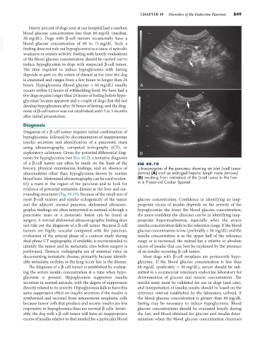
of a β-cell tumor can often be made on the basis of the FIG 49.19
history, physical examination findings, and an absence of Ultrasonogram of the pancreas showing an islet β-cell tumor
abnormalities other than hypoglycemia shown by routine (arrow) (A) and an enlarged hepatic lymph node (arrows)
blood tests. Abdominal ultrasonography can be used to iden- (B) resulting from metastasis of the β-cell tumor to the liver
tify a mass in the region of the pancreas and to look for in a 9-year-old Cocker Spaniel.
evidence of potential metastatic disease in the liver and sur-
rounding structures (Fig. 49.19). Because of the small size of
most β-cell tumors and similar echogenicity of the tumor glucose concentration. Confidence in identifying an inap-
and the adjacent normal pancreas, abdominal ultrasono- propriate excess of insulin depends on the severity of the
graphic findings are often interpreted as normal, although a hypoglycemia; the lower the blood glucose concentration,
pancreatic mass or a metastatic lesion can be found at the more confident the clinician can be in identifying inap-
surgery. A normal abdominal ultrasonographic finding does propriate hyperinsulinemia, especially when the serum
not rule out the diagnosis of a β-cell tumor. Because β-cell insulin concentration falls in the reference range. If the blood
tumors are highly vascular compared with the pancreas, glucose concentration is low (preferably < 50 mg/dL) and the
evaluation of the arterial phase of a contrast study during insulin concentration is in the upper half of the reference
dual-phase CT angiography, if available, is recommended to range or is increased, the animal has a relative or absolute
identify the tumor and its metastatic sites before surgery is excess of insulin that can best be explained by the presence
performed. Thoracic radiographs are of minimal value in of an insulin-secreting β-cell tumor.
documenting metastatic disease, primarily because identifi- Most dogs with β-cell neoplasia are persistently hypo-
able metastatic nodules in the lung occur late in the disease. glycemic. If the blood glucose concentration is less than
The diagnosis of a β-cell tumor is established by evaluat- 60 mg/dL (preferably < 50 mg/dL), serum should be sub-
ing the serum insulin concentration at a time when hypo- mitted to a commercial veterinary endocrine laboratory for
glycemia is present. Hypoglycemia suppresses insulin determination of glucose and insulin concentration. The
secretion in normal animals, with the degree of suppression insulin assay must be validated for use in dogs (and cats),
directly related to its severity. Hypoglycemia fails to have this and interpretation of insulin results should be based on the
same suppressive effect on insulin secretion if the insulin is reference interval established by the laboratory utilized. If
synthesized and secreted from autonomous neoplastic cells the blood glucose concentration is greater than 60 mg/dL,
because tumor cells that produce and secrete insulin are less fasting may be necessary to induce hypoglycemia. Blood
responsive to hypoglycemia than are normal β cells. Invari- glucose concentrations should be evaluated hourly during
ably, the dog with a β-cell tumor will have an inappropriate the fast, and blood obtained for glucose and insulin deter-
excess of insulin relative to that needed for a particular blood mination when the blood glucose concentration decreases