
Page 881 - Small Animal Internal Medicine, 6th Edition
P. 881
CHAPTER 49 Disorders of the Endocrine Pancreas 853
Prognosis
The long-term prognosis for β-cell neoplasia is guarded to BOX 49.15
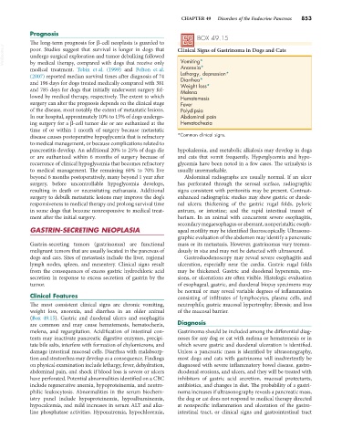
VetBooks.ir poor. Studies suggest that survival is longer in dogs that Clinical Signs of Gastrinoma in Dogs and Cats
undergo surgical exploration and tumor debulking followed
Vomiting*
by medical therapy, compared with dogs that receive only
medical treatment. Tobin et al. (1999) and Polton et al. Anorexia*
(2007) reported median survival times after diagnosis of 74 Lethargy, depression*
Diarrhea*
and 196 days for dogs treated medically compared with 381 Weight loss*
and 785 days for dogs that initially underwent surgery fol- Melena
lowed by medical therapy, respectively. The extent to which Hematemesis
surgery can alter the prognosis depends on the clinical stage Fever
of the disease, most notably the extent of metastatic lesions. Polydipsia
In our hospital, approximately 10% to 15% of dogs undergo- Abdominal pain
ing surgery for a β-cell tumor die or are euthanized at the Hematochezia
time of or within 1 month of surgery because metastatic
disease causes postoperative hypoglycemia that is refractory *Common clinical signs.
to medical management, or because complications related to
pancreatitis develop. An additional 20% to 25% of dogs die hypokalemia, and metabolic alkalosis may develop in dogs
or are euthanized within 6 months of surgery because of and cats that vomit frequently. Hyperglycemia and hypo-
recurrence of clinical hypoglycemia that becomes refractory glycemia have been noted in a few cases. The urinalysis is
to medical management. The remaining 60% to 70% live usually unremarkable.
beyond 6 months postoperatively, many beyond 1 year after Abdominal radiographs are usually normal. If an ulcer
surgery, before uncontrollable hypoglycemia develops, has perforated through the serosal surface, radiographic
resulting in death or necessitating euthanasia. Additional signs consistent with peritonitis may be present. Contrast-
surgery to debulk metastatic lesions may improve the dog’s enhanced radiographic studies may show gastric or duode-
responsiveness to medical therapy and prolong survival time nal ulcers; thickening of the gastric rugal folds, pyloric
in some dogs that become nonresponsive to medical treat- antrum, or intestine; and the rapid intestinal transit of
ment after the initial surgery. barium. In an animal with concurrent severe esophagitis,
secondary megaesophagus or aberrant, nonperistaltic esoph-
GASTRIN-SECRETING NEOPLASIA ageal motility may be identified fluoroscopically. Ultrasono-
graphic evaluation of the abdomen may identify a pancreatic
Gastrin-secreting tumors (gastrinomas) are functional mass or its metastasis. However, gastrinomas vary tremen-
malignant tumors that are usually located in the pancreas of dously in size and may not be detected with ultrasound.
dogs and cats. Sites of metastasis include the liver, regional Gastroduodenoscopy may reveal severe esophagitis and
lymph nodes, spleen, and mesentery. Clinical signs result ulceration, especially near the cardia. Gastric rugal folds
from the consequences of excess gastric hydrochloric acid may be thickened. Gastric and duodenal hyperemia, ero-
secretion in response to excess secretion of gastrin by the sions, or ulcerations are often visible. Histologic evaluation
tumor. of esophageal, gastric, and duodenal biopsy specimens may
be normal or may reveal variable degrees of inflammation
Clinical Features consisting of infiltrates of lymphocytes, plasma cells, and
The most consistent clinical signs are chronic vomiting, neutrophils; gastric mucosal hypertrophy; fibrosis; and loss
weight loss, anorexia, and diarrhea in an older animal of the mucosal barrier.
(Box 49.15). Gastric and duodenal ulcers and esophagitis
are common and may cause hematemesis, hematochezia, Diagnosis
melena, and regurgitation. Acidification of intestinal con- Gastrinoma should be included among the differential diag-
tents may inactivate pancreatic digestive enzymes, precipi- noses for any dog or cat with melena or hematemesis or in
tate bile salts, interfere with formation of chylomicrons, and which severe gastric and duodenal ulceration is identified.
damage intestinal mucosal cells. Diarrhea with malabsorp- Unless a pancreatic mass is identified by ultrasonography,
tion and steatorrhea may develop as a consequence. Findings most dogs and cats with gastrinoma will inadvertently be
on physical examination include lethargy, fever, dehydration, diagnosed with severe inflammatory bowel disease, gastro-
abdominal pain, and shock if blood loss is severe or ulcers duodenal erosions, and ulcers, and they will be treated with
have perforated. Potential abnormalities identified on a CBC inhibitors of gastric acid secretion, mucosal protectants,
include regenerative anemia, hypoproteinemia, and neutro- antibiotics, and changes in diet. The probability of a gastri-
philic leukocytosis. Abnormalities in the serum biochem- noma increases if ultrasonography reveals a pancreatic mass,
istry panel include hypoproteinemia, hypoalbuminemia, the dog or cat does not respond to medical therapy directed
hypocalcemia, and mild increases in serum ALT and alka- at nonspecific inflammation and ulceration of the gastro-
line phosphatase activities. Hyponatremia, hypochloremia, intestinal tract, or clinical signs and gastrointestinal tract