
Page 886 - Small Animal Internal Medicine, 6th Edition
P. 886
858 PART VI Endocrine Disorders
VetBooks.ir
A B
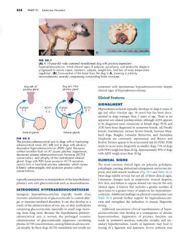
FIG 50.1
(A) A 10-year-old male castrated mixed-breed dog with pituitary-dependent
hyperadrenocorticism. Initial clinical signs of polyuria, polydipsia, and endocrine alopecia
progressed to severe stupor, anorexia, adipsia, weight loss, and loss of body temperature
regulation. (B) Cross-section of the brain from the dog in A, showing a pituitary
macroadenoma severely compressing surrounding brain structures.
Dog with AT Dog with PDH consistent with spontaneous hypoadrenocorticism despite
pituitary gland pituitary gland clinical signs of hyperadrenocorticism.
Clinical Features
SIGNALMENT
Cortisol ACTH Cortisol ACTH Hyperadrenocorticism typically develops in dogs 6 years of
age and older (median age, 10 years) but has been docu-
mented in dogs younger than 5 years of age. There is no
apparent sex-related predisposition, although ADH appears
to be diagnosed more commonly in female dogs. PDH and
ATH have been diagnosed in numerous breeds. All Poodle
breeds, Dachshunds, various Terrier breeds, German Shep-
Adrenal glands Adrenal glands
herd dogs, Beagles, Labrador Retrievers, and Australian
FIG 50.2 Shepherds are commonly represented, and Boxers and
The pituitary-adrenocortical axis in dogs with a functioning Boston Terriers appear to be at increased risk for PDH. PDH
adrenocortical tumor (AT; left) and in dogs with pituitary- tends to occur more frequently in smaller dogs; 75% of dogs
dependent hyperadrenocorticism (PDH; right). Excessive
cortisol secretion from an AT causes pituitary suppression, with PDH weigh less than 20 kg. Approximately 50% of dogs
decreased plasma adrenocorticotropic hormone (ACTH) with ADH weigh more than 20 kg.
concentration, and atrophy of the contralateral adrenal
gland. Dogs with PDH have excessive ACTH secretion, CLINICAL SIGNS
usually from a functional pituitary adenoma, which causes The most common clinical signs are polyuria, polydipsia,
bilateral adrenomegaly and excessive plasma cortisol polyphagia, panting, abdominal enlargement, endocrine alo-
concentrations. pecia, and mild muscle weakness (Fig. 50.3 and Table 50.1).
Most dogs exhibit several, but not all, of these clinical signs.
typically unresponsive to manipulation of the hypothalamic- Cutaneous changes such as nonpruritic truncal alopecia,
pituitary axis with glucocorticoids such as dexamethasone. thin skin, and failure to regrow shaved hair may be the only
clinical signs. A history that includes a greater number of
IATROGENIC HYPERADRENOCORTICISM signs leads to a greater index of suspicion for hyperadreno-
Iatrogenic hyperadrenocorticism typically results from corticism. Additional findings on physical examination (see
excessive administration of glucocorticoids to control aller- Table 50.1) provide further support for hyperadrenocorti-
gic or immune-mediated disorders. It can also develop as a cism and strengthen the indication to pursue diagnostic
result of the administration of eye, ear, or skin medications testing.
containing glucocorticoids, especially in small dogs receiv- Additional uncommon clinical manifestations of hyper-
ing them long term. Because the hypothalamic-pituitary- adrenocorticism may develop as a consequence of chronic
adrenocortical axis is normal, the prolonged excessive hypercortisolism. Suppression of pituitary function can
administration of glucocorticoids suppresses circulating result in persistent anestrus, testicular atrophy, and sec-
plasma ACTH concentrations, causing bilateral adrenocorti- ondary hypothyroidism. Laxity of ligaments may lead to
cal atrophy. In these dogs ACTH stimulation test results are tearing of a ligament and lameness. Severe polyuria may