
Page 889 - Small Animal Internal Medicine, 6th Edition
P. 889
CHAPTER 50 Disorders of the Adrenal Gland 861
hypercoagulability, which is a common finding at the time BOX 50.2
hyperadrenocorticism is diagnosed, may persist despite
VetBooks.ir treatment of PDH with trilostane, and may result in sponta- Clinicopathologic Abnormalities Commonly Identified in
neous thromboembolism, typically involving pulmonary
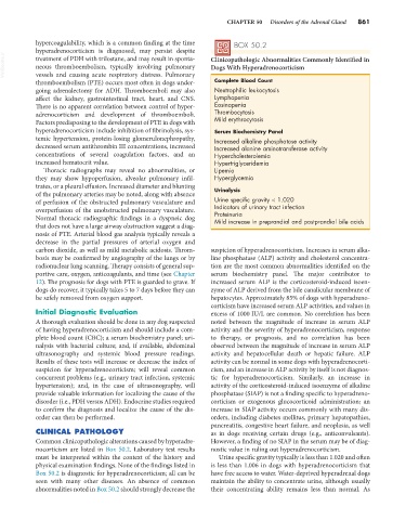
Dogs With Hyperadrenocorticism
vessels and causing acute respiratory distress. Pulmonary
thromboembolism (PTE) occurs most often in dogs under- Complete Blood Count
going adrenalectomy for ADH. Thromboemboli may also Neutrophilic leukocytosis
affect the kidney, gastrointestinal tract, heart, and CNS. Lymphopenia
There is no apparent correlation between control of hyper- Eosinopenia
adrenocorticism and development of thromboemboli. Thrombocytosis
Factors predisposing to the development of PTE in dogs with Mild erythrocytosis
hyperadrenocorticism include inhibition of fibrinolysis, sys- Serum Biochemistry Panel
temic hypertension, protein-losing glomerulonephropathy, Increased alkaline phosphatase activity
decreased serum antithrombin III concentrations, increased Increased alanine aminotransferase activity
concentrations of several coagulation factors, and an Hypercholesterolemia
increased hematocrit value. Hypertriglyceridemia
Thoracic radiographs may reveal no abnormalities, or Lipemia
they may show hypoperfusion, alveolar pulmonary infil- Hyperglycemia
trates, or a pleural effusion. Increased diameter and blunting Urinalysis
of the pulmonary arteries may be noted, along with absence
of perfusion of the obstructed pulmonary vasculature and Urine specific gravity < 1.020
overperfusion of the unobstructed pulmonary vasculature. Indicators of urinary tract infection
Proteinuria
Normal thoracic radiographic findings in a dyspneic dog Mild increase in preprandial and postprandial bile acids
that does not have a large airway obstruction suggest a diag-
nosis of PTE. Arterial blood gas analysis typically reveals a
decrease in the partial pressures of arterial oxygen and
carbon dioxide, as well as mild metabolic acidosis. Throm- suspicion of hyperadrenocorticism. Increases in serum alka-
bosis may be confirmed by angiography of the lungs or by line phosphatase (ALP) activity and cholesterol concentra-
radionuclear lung scanning. Therapy consists of general sup- tion are the most common abnormalities identified on the
portive care, oxygen, anticoagulants, and time (see Chapter serum biochemistry panel. The major contributor to
12). The prognosis for dogs with PTE is guarded to grave. If increased serum ALP is the corticosteroid-induced isoen-
dogs do recover, it typically takes 5 to 7 days before they can zyme of ALP derived from the bile canalicular membrane of
be safely removed from oxygen support. hepatocytes. Approximately 85% of dogs with hyperadreno-
corticism have increased serum ALP activities, and values in
Initial Diagnostic Evaluation excess of 1000 IU/L are common. No correlation has been
A thorough evaluation should be done in any dog suspected noted between the magnitude of increase in serum ALP
of having hyperadrenocorticism and should include a com- activity and the severity of hyperadrenocorticism, response
plete blood count (CBC); a serum biochemistry panel; uri- to therapy, or prognosis, and no correlation has been
nalysis with bacterial culture; and, if available, abdominal observed between the magnitude of increase in serum ALP
ultrasonography and systemic blood pressure readings. activity and hepatocellular death or hepatic failure. ALP
Results of these tests will increase or decrease the index of activity can be normal in some dogs with hyperadrenocorti-
suspicion for hyperadrenocorticism; will reveal common cism, and an increase in ALP activity by itself is not diagnos-
concurrent problems (e.g., urinary tract infection, systemic tic for hyperadrenocorticism. Similarly, an increase in
hypertension); and, in the case of ultrasonography, will activity of the corticosteroid-induced isoenzyme of alkaline
provide valuable information for localizing the cause of the phosphatase (SIAP) is not a finding specific to hyperadreno-
disorder (i.e., PDH versus ADH). Endocrine studies required corticism or exogenous glucocorticoid administration; an
to confirm the diagnosis and localize the cause of the dis- increase in SIAP activity occurs commonly with many dis-
order can then be performed. orders, including diabetes mellitus, primary hepatopathies,
pancreatitis, congestive heart failure, and neoplasia, as well
CLINICAL PATHOLOGY as in dogs receiving certain drugs (e.g., anticonvulsants).
Common clinicopathologic alterations caused by hyperadre- However, a finding of no SIAP in the serum may be of diag-
nocorticism are listed in Box 50.2. Laboratory test results nostic value in ruling out hyperadrenocorticism.
must be interpreted within the context of the history and Urine specific gravity typically is less than 1.020 and often
physical examination findings. None of the findings listed in is less than 1.006 in dogs with hyperadrenocorticism that
Box 50.2 is diagnostic for hyperadrenocorticism; all can be have free access to water. Water-deprived hyperadrenal dogs
seen with many other diseases. An absence of common maintain the ability to concentrate urine, although usually
abnormalities noted in Box 50.2 should strongly decrease the their concentrating ability remains less than normal. As