
Page 890 - Small Animal Internal Medicine, 6th Edition
P. 890
862 PART VI Endocrine Disorders
such, urine specific gravities of 1.020 to 1.025 may be identi- BOX 50.3
fied if urine is obtained after water has been withheld from
VetBooks.ir the dog. Abnormalities Identified by Abdominal and Thoracic
Proteinuria is a common finding in dogs with untreated
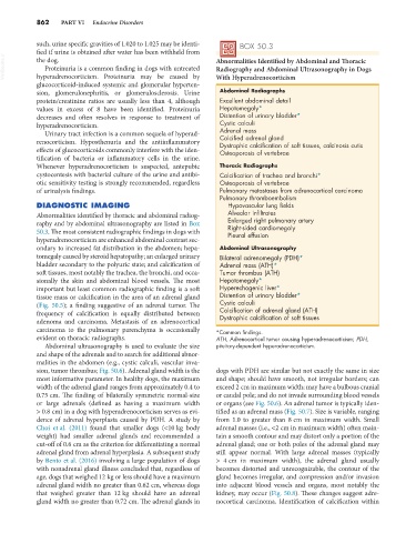
Radiography and Abdominal Ultrasonography in Dogs
hyperadrenocorticism. Proteinuria may be caused by
glucocorticoid-induced systemic and glomerular hyperten- With Hyperadrenocorticism
sion, glomerulonephritis, or glomerulosclerosis. Urine Abdominal Radiographs
protein/creatinine ratios are usually less than 4, although Excellent abdominal detail
values in excess of 8 have been identified. Proteinuria Hepatomegaly*
decreases and often resolves in response to treatment of Distention of urinary bladder*
hyperadrenocorticism. Cystic calculi
Urinary tract infection is a common sequela of hyperad- Adrenal mass
renocorticism. Hyposthenuria and the antiinflammatory Calcified adrenal gland
Dystrophic calcification of soft tissues, calcinosis cutis
effects of glucocorticoids commonly interfere with the iden- Osteoporosis of vertebrae
tification of bacteria or inflammatory cells in the urine.
Whenever hyperadrenocorticism is suspected, antepubic Thoracic Radiographs
cystocentesis with bacterial culture of the urine and antibi- Calcification of trachea and bronchi*
otic sensitivity testing is strongly recommended, regardless Osteoporosis of vertebrae
of urinalysis findings. Pulmonary metastases from adrenocortical carcinoma
Pulmonary thromboembolism
DIAGNOSTIC IMAGING Hypovascular lung fields
Abnormalities identified by thoracic and abdominal radiog- Alveolar infiltrates
raphy and by abdominal ultrasonography are listed in Box Enlarged right pulmonary artery
Right-sided cardiomegaly
50.3. The most consistent radiographic findings in dogs with Pleural effusion
hyperadrenocorticism are enhanced abdominal contrast sec-
ondary to increased fat distribution in the abdomen; hepa- Abdominal Ultrasonography
tomegaly caused by steroid hepatopathy; an enlarged urinary Bilateral adrenomegaly (PDH)*
bladder secondary to the polyuric state; and calcification of Adrenal mass (ATH)*
soft tissues, most notably the trachea, the bronchi, and occa- Tumor thrombus (ATH)
sionally the skin and abdominal blood vessels. The most Hepatomegaly*
important but least common radiographic finding is a soft Hyperechogenic liver*
tissue mass or calcification in the area of an adrenal gland Distention of urinary bladder*
(Fig. 50.5); a finding suggestive of an adrenal tumor. The Cystic calculi
frequency of calcification is equally distributed between Calcification of adrenal gland (ATH)
Dystrophic calcification of soft tissues
adenoma and carcinoma. Metastasis of an adrenocortical
carcinoma to the pulmonary parenchyma is occasionally *Common findings.
evident on thoracic radiographs. ATH, Adrenocortical tumor causing hyperadrenocorticism; PDH,
Abdominal ultrasonography is used to evaluate the size pituitary-dependent hyperadrenocorticism.
and shape of the adrenals and to search for additional abnor-
malities in the abdomen (e.g., cystic calculi, vascular inva-
sion, tumor thrombus; Fig. 50.6). Adrenal gland width is the dogs with PDH are similar but not exactly the same in size
most informative parameter. In healthy dogs, the maximum and shape; should have smooth, not irregular borders; can
width of the adrenal gland ranges from approximately 0.4 to exceed 2 cm in maximum width; may have a bulbous cranial
0.75 cm. The finding of bilaterally symmetric normal-size or caudal pole; and do not invade surrounding blood vessels
or large adrenals (defined as having a maximum width or organs (see Fig. 50.6). An adrenal tumor is typically iden-
> 0.8 cm) in a dog with hyperadrenocorticism serves as evi- tified as an adrenal mass (Fig. 50.7). Size is variable, ranging
dence of adrenal hyperplasia caused by PDH. A study by from 1.0 to greater than 8 cm in maximum width. Small
Choi et al. (2011) found that smaller dogs (<10 kg body adrenal masses (i.e., <2 cm in maximum width) often main-
weight) had smaller adrenal glands and recommended a tain a smooth contour and may distort only a portion of the
cut-off of 0.6 cm as the criterion for differentiating a normal adrenal gland; one or both poles of the adrenal gland may
adrenal gland from adrenal hyperplasia. A subsequent study still appear normal. With large adrenal masses (typically
by Bento et al. (2016) involving a large population of dogs > 4 cm in maximum width), the adrenal gland usually
with nonadrenal gland illness concluded that, regardless of becomes distorted and unrecognizable, the contour of the
age, dogs that weighed 12 kg or less should have a maximum gland becomes irregular, and compression and/or invasion
adrenal gland width no greater than 0.62 cm, whereas dogs into adjacent blood vessels and organs, most notably the
that weighed greater than 12 kg should have an adrenal kidney, may occur (Fig. 50.8). These changes suggest adre-
gland width no greater than 0.72 cm. The adrenal glands in nocortical carcinoma. Identification of calcification within