
Page 10 - Veterinary diagnostic imaging birds exotic pets wildlife
P. 10
6 SECTION I III The Birds
III WILD BIRDS: DIAGNOSTIC intensity lamp (Figure 1-7). A VD image of a medium-
QUALITY—THE PRIME DIRECTIVE sized raptor on a large film with the wings partially
extended should be sufficiently symmetrical to allow
Diagnostic quality depends on two essential ingredi- for a close comparison between the injured and normal
ents: (1) appearance and (2) positioning. Appearance wings (Figure 1-8).
refers to image contrast and detail, and positioning
refers to how well a standard projection approaches
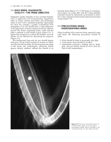
the ideal. For example, a quality ventrodorsal (VD)
radiograph of an owl should show a clear difference III PRECAUTIONS WHEN
in the appearance of bones compared with the adjacent RADIOGRAPHING BIRDS
muscle: the former appearing nearly white and the
latter a medium to dark shade of gray (Figure 1-5). A When handling fully conscious birds, especially large
lighter fi lm designed to evaluate the feathers and soft wild birds, the following precautions should be
tissues can be obtained by decreasing the exposure taken:
(Figure 1-6).
The background lung and air sacs should appear • Work should be done in reasonably dim light.
dark gray, except where overlain by muscle, in which • Unnecessary noise should be avoided.
case they become invisible. For the most part, the edges • Appropriate protective clothing, gloves, head-
of the bones and cardiohepatic silhouette should gear, and eye shields should be worn until the
appear sharply outlined without the benefit of an bird is fully incapacitated.
Figure 1-5 • Close-up ventrodorsal view of
the right elbow region of an owl with a
large-gauge shotgun pellet embedded in the
muscles between its right radius and ulna.
2/11/2008 10:50:37 AM
ch001-A02527.indd 6 2/11/2008 10:50:37 AM
ch001-A02527.indd 6