
Page 12 - Veterinary diagnostic imaging birds exotic pets wildlife
P. 12
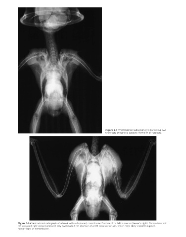
Figure 1-7 • Ventrodorsal radiograph of a burrowing owl
                                                                               under gas anesthesia appears normal in all respects.


                    Figure 1-8 • Ventrodorsal radiograph of a hawk with a displaced, comminuted fracture of its left humerus (viewer’s right). Comparison with
                    the uninjured right wing reveals not only swelling but the absence of a left clavicular air sac, which most likely indicates rupture,
                    hemorrhage, or compression.


                                                                                                                      2/11/2008   10:50:38 AM
           ch001-A02527.indd   8                                                                                      2/11/2008   10:50:38 AM
           ch001-A02527.indd   8