
Page 113 - Veterinary diagnostic imaging birds exotic pets wildlife
P. 113
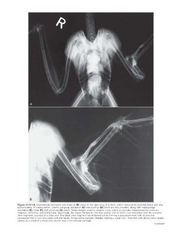
A


                             B

                    Figure 10-15 • A, Ventrodorsal orientation and close-up (B) views of the right wing of a hawk, which fractured its proximal radius and ulna
                    approximately 10 weeks earlier. Lateral, wings-up orientation (C) and close-up (D) views are also provided, along with leading-edge
                    orientation (E), close (F), and ultraclose (G) views. These images reveal a complex injury, which is currently characterized by nonunion,

                    malunion, deformity, and dysfunction. Specifically, the radius has broken into four pieces, one of which now articulates with the proximal
                    ulnar fragment, courtesy of a false joint. The distal ulnar fragment has followed suit by forming a pseudoarthrosis with its proximal
                    counterpart that in turn articulates with the newly formed radioulnar joint, thereby creating a super joint. Note that both elbow joints appear
                    narrow as a result of a temporary volume loss in the articular cartilage.
                                                                                                                  Continued


                                                                                                                      2/11/2008   10:56:19 AM
           ch010-A02527.indd   109
           ch010-A02527.indd   109                                                                                    2/11/2008   10:56:19 AM