
Page 168 - Veterinary diagnostic imaging birds exotic pets wildlife
P. 168
164 SECTION I III The Birds
A
B
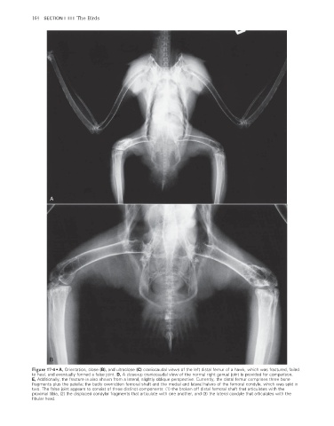
Figure 17-4 • A, Orientation, close (B), and ultraclose (C) craniocaudal views of the left distal femur of a hawk, which was fractured, failed
to heal, and eventually formed a false joint. D, A close-up craniocaudal view of the normal right genual joint is provided for comparison.
E, Additionally, the fracture is also shown from a lateral, slightly oblique perspective. Currently, the distal femur comprises three bone
fragments plus the patella: the badly overridden femoral shaft and the medial and lateral halves of the femoral condyle, which was split in
two. The false joint appears to consist of three distinct components: (1) the broken off distal femoral shaft that articulates with the
proximal tibia, (2) the displaced condylar fragments that articulate with one another, and (3) the lateral condyle that articulates with the
fi bular head.
2/11/2008 11:00:05 AM
ch017-A02527.indd 164 2/11/2008 11:00:05 AM
ch017-A02527.indd 164