
Page 170 - Veterinary diagnostic imaging birds exotic pets wildlife
P. 170
166 SECTION I III The Birds
A B
C
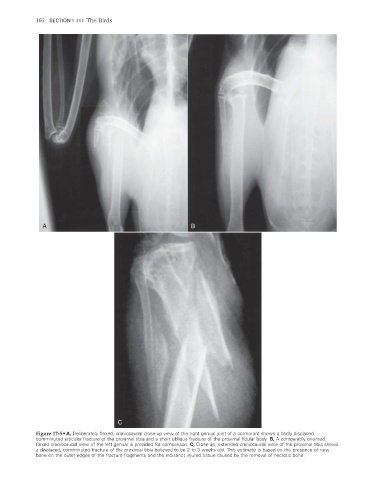
Figure 17-5 • A, Deliberately flexed, craniocaudal close-up view of the right genual joint of a cormorant shows a badly displaced,
comminuted articular fracture of the proximal tibia and a short oblique fracture of the proximal fi bular body. B, A comparably oriented,
fl exed craniocaudal view of the left genual is provided for comparison. C, Close-up, extended craniocaudal view of the proximal tibia shows
a displaced, comminuted fracture of the proximal tibia believed to be 2 to 3 weeks old. This estimate is based on the presence of new
bone on the outer edges of the fracture fragments and the indistinct injured tissue caused by the removal of necrotic bone.
2/11/2008 11:00:09 AM
ch017-A02527.indd 166 2/11/2008 11:00:09 AM
ch017-A02527.indd 166