
Page 193 - Veterinary diagnostic imaging birds exotic pets wildlife
P. 193
CHAPTER 18 III The Head 189
Facial Region
The facial region of birds features the eyes, which are
preeminent in some species such as raptors (Figures
18-17 and 18-18). Radiographically, the eyes appear as
large circular densities, nearly as large as the adjacent
brain (Figure 18-19).
Birds also have facial sinuses, which are quite elabo-
rate in parrots and related birds. The primary sinus
chamber, the infraorbital sinus, encircles the ventral half
of the obit and then extends outward around the eyes
and ears in an elaborate system of irregular channels,
or diverticula. Some of these channels reach as far
forward as the central conchae and mandible and as
far caudally as the neck in the form of the cervicoce-
phalic air sac.
Most of this intricate system of air-fi lled space is
difficult or impossible to identify as individual struc-
ture, but it is possible to identify some of the larger
elements such as the rostral portion of the infraorbital
sinus, which appears as a triangular lucency immedi-
ately forward of the eye, as seen in a lateral projection
of the head (Figures 18-20 through 18-24).
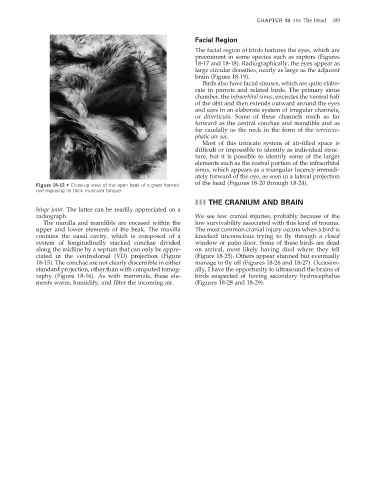
Figure 18-12 • Close-up view of the open beak of a great horned
owl exposing its thick muscular tongue.
III THE CRANIUM AND BRAIN
hinge joint. The latter can be readily appreciated on a
radiograph. We see few cranial injuries, probably because of the
The maxilla and mandible are encased within the low survivability associated with this kind of trauma.
upper and lower elements of the beak. The maxilla The most common cranial injury occurs when a bird is
contains the nasal cavity, which is composed of a knocked unconscious trying to fly through a closed
system of longitudinally stacked conchae divided window or patio door. Some of these birds are dead
along the midline by a septum that can only be appre- on arrival, most likely having died where they fell
ciated in the ventrodorsal (VD) projection (Figure (Figure 18-25). Others appear stunned but eventually
18-15). The conchae are not clearly discernible in either manage to fly off (Figures 18-26 and 18-27). Occasion-
standard projection, other than with computed tomog- ally, I have the opportunity to ultrasound the brains of
raphy (Figure 18-16). As with mammals, these ele- birds suspected of having secondary hydrocephalus
ments warm, humidify, and fi lter the incoming air. (Figures 18-28 and 18-29).
2/11/2008 11:01:04 AM
ch018-A02527.indd 189
ch018-A02527.indd 189 2/11/2008 11:01:04 AM