Page 195 - Veterinary diagnostic imaging birds exotic pets wildlife
P. 195

CHAPTER 18  III The Head  191


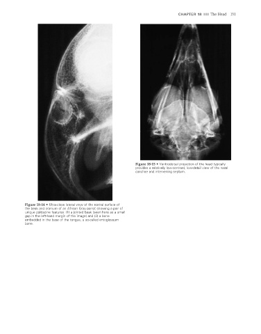
                                                                         Figure 18-15  •  Ventrodorsal projection of the head typically
                                                                         provides a relatively low-contrast, low-detail view of the nasal
                                                                         conchae and intervening septum.


                    Figure 18-14  •  Ultra-close lateral view of the rostral surface of
                    the beak and cranium of an African Gray parrot showing a pair of
                    unique psittacine features: (1) a jointed beak (seen here as a small
                    gap in the left-hand margin of the image) and (2) a bone
                    embedded in the base of the tongue, a so-called entoglossum
                    bone.


                                                                                                                      2/11/2008   11:01:06 AM
           ch018-A02527.indd   191                                                                                    2/11/2008   11:01:06 AM
           ch018-A02527.indd   191
   190   191   192   193   194   195   196   197   198   199   200