Page 208 - Veterinary diagnostic imaging birds exotic pets wildlife
P. 208
204  SECTION I  III  The Birds


                             A


                                                B
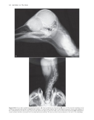
                    Figure 19-4  •  Crow with a partial esophageal obstruction. A, Close-up lateral and ventrodorsal (B) views of the proximal esophagus show
                    moderate localized swelling that is displacing the trachea in a ventromedial direction. C, A ventrolateral sonogram shows what appears to

                    be a well-defined accumulation of pus with air at its center and along its perimeter. D, Close-up lateral and ventrodorsal (E) esophagrams
                    reveal contrast retention coincident with the aforementioned swelling. The lesion proved to be an abscess in the wall of the esophagus.


                                                                                                                      2/11/2008   11:08:13 AM
           ch019-A02527.indd   204                                                                                    2/11/2008   11:08:13 AM
           ch019-A02527.indd   204

