
Page 211 - Veterinary diagnostic imaging birds exotic pets wildlife
P. 211
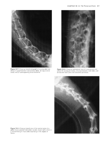
CHAPTER 19  III  The Throat and Neck  207


                    Figure 19-7  •  Close-up lateral radiograph of a young adult owl   Figure 19-8  •  Close-up ventrodorsal view of a young adult owl
                    shows (1) small indistinct intervertebral disks, (2) vague spinal   shows (1) rectangular bodies, (2) proportionately sized disks, and
                    canal, and (3) fused-appearing dorsal elements.      (3) discrete facet joints and transverse processes.


                    Figure 19-9  •  Close-up lateral view of the cervical spine of a
                    grebe shows apparent dislocation caudally (lower right), which is
                    a normal finding in many birds depending on the degree of

                    fl exion.


                                                                                                                      2/11/2008   11:08:17 AM
           ch019-A02527.indd   207                                                                                    2/11/2008   11:08:17 AM
           ch019-A02527.indd   207