
Page 214 - Veterinary diagnostic imaging birds exotic pets wildlife
P. 214
210 SECTION I III The Birds
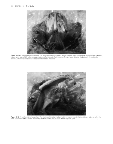
Figure 20-1 • Great horned owl dissection. The bird is positioned on its back, and its pectoral and supracoracoideus muscles are laid open,
exposing the keel, which is the principal attachment for the major fl ight muscles. The V-shaped object at the bottom is formed by the
clavicles, which in some species is fused and termed the wishbone.
Figure 20-2 • Great horned owl dissection. The bird is positioned on its back but is now seen from in front and to one side, revealing the
distal attachment of the coracoid (lower left), the keel (center), and much of the rib cage (far right).
2/11/2008 11:08:42 AM
ch020-A02527.indd 210
ch020-A02527.indd 210 2/11/2008 11:08:42 AM