
Page 215 - Veterinary diagnostic imaging birds exotic pets wildlife
P. 215
CHAPTER 20 III The Torso 211
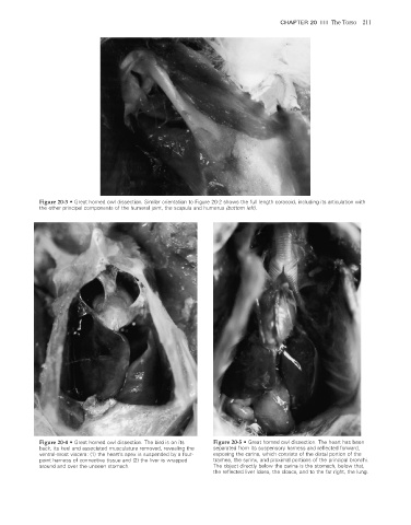
Figure 20-3 • Great horned owl dissection. Similar orientation to Figure 20-2 shows the full length coracoid, including its articulation with
the other principal components of the humeral joint, the scapula and humerus (bottom left).
Figure 20-4 • Great horned owl dissection. The bird is on its Figure 20-5 • Great horned owl dissection. The heart has been
back, its keel and associated musculature removed, revealing the separated from its suspensory harness and refl ected forward,
ventral-most viscera: (1) the heart’s apex is suspended by a four- exposing the carina, which consists of the distal portion of the
point harness of connective tissue and (2) the liver is wrapped trachea, the syrinx, and proximal portions of the principal bronchi.
around and over the unseen stomach. The object directly below the carina is the stomach, below that,
the reflected liver lobes, the cloaca, and to the far right, the lung.
2/11/2008 11:08:42 AM
ch020-A02527.indd 211 2/11/2008 11:08:42 AM
ch020-A02527.indd 211