
Page 220 - Veterinary diagnostic imaging birds exotic pets wildlife
P. 220
216 SECTION I III The Birds
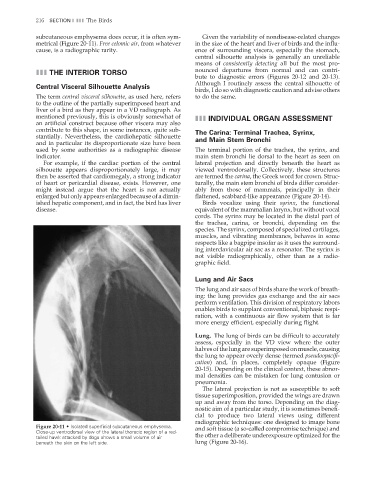
subcutaneous emphysema does occur, it is often sym- Given the variability of nondisease-related changes
metrical (Figure 20-11). Free celomic air, from whatever in the size of the heart and liver of birds and the infl u-
cause, is a radiographic rarity. ence of surrounding viscera, especially the stomach,
central silhouette analysis is generally an unreliable
means of consistently detecting all but the most pro-
III THE INTERIOR TORSO nounced departures from normal and can contri-
bute to diagnostic errors (Figures 20-12 and 20-13).
Although I routinely assess the central silhouette of
Central Visceral Silhouette Analysis
birds, I do so with diagnostic caution and advise others
The term central visceral silhouette, as used here, refers to do the same.
to the outline of the partially superimposed heart and
liver of a bird as they appear in a VD radiograph. As
mentioned previously, this is obviously somewhat of III INDIVIDUAL ORGAN ASSESSMENT
an artificial construct because other viscera may also
contribute to this shape, in some instances, quite sub- The Carina: Terminal Trachea, Syrinx,
stantially. Nevertheless, the cardiohepatic silhouette and Main Stem Bronchi
and in particular its disproportionate size have been
used by some authorities as a radiographic disease The terminal portion of the trachea, the syrinx, and
indicator. main stem bronchi lie dorsal to the heart as seen on
For example, if the cardiac portion of the central lateral projection and directly beneath the heart as
silhouette appears disproportionately large, it may viewed ventrodorsally. Collectively, these structures
then be asserted that cardiomegaly, a strong indicator are termed the carina, the Greek word for crown. Struc-
of heart or pericardial disease, exists. However, one turally, the main stem bronchi of birds differ consider-
might instead argue that the heart is not actually ably from those of mammals, principally in their
enlarged but only appears enlarged because of a dimin- fl attened, scabbard-like appearance (Figure 20-14).
ished hepatic component, and in fact, the bird has liver Birds vocalize using their syrinx, the functional
disease. equivalent of the mammalian larynx, but without vocal
cords. The syrinx may be located in the distal part of
the trachea, carina, or bronchi, depending on the
species. The syrinx, composed of specialized cartilages,
muscles, and vibrating membranes, behaves in some
respects like a bagpipe insofar as it uses the surround-
ing interclavicular air sac as a resonator. The syrinx is
not visible radiographically, other than as a radio-
graphic fi eld.
Lung and Air Sacs
The lung and air sacs of birds share the work of breath-
ing: the lung provides gas exchange and the air sacs
perform ventilation. This division of respiratory labors
enables birds to supplant conventional, biphasic respi-
ration, with a continuous air flow system that is far
more energy effi cient, especially during fl ight.
Lung. The lung of birds can be difficult to accurately
assess, especially in the VD view where the outer
halves of the lung are superimposed on muscle, causing
the lung to appear overly dense (termed pseudoopacifi -
cation) and, in places, completely opaque (Figure
20-15). Depending on the clinical context, these abnor-
mal densities can be mistaken for lung contusion or
pneumonia.
The lateral projection is not as susceptible to soft
tissue superimposition, provided the wings are drawn
up and away from the torso. Depending on the diag-
nostic aim of a particular study, it is sometimes benefi -
cial to produce two lateral views using different
radiographic techniques: one designed to image bone
Figure 20-11 • Isolated superfi cial subcutaneous emphysema. and soft tissue (a so-called compromise technique) and
Close-up ventrodorsal view of the lateral thoracic region of a red-
tailed hawk attacked by dogs shows a small volume of air the other a deliberate underexposure optimized for the
beneath the skin on the left side. lung (Figure 20-16).
2/11/2008 11:08:48 AM
ch020-A02527.indd 216 2/11/2008 11:08:48 AM
ch020-A02527.indd 216