
Page 224 - Veterinary diagnostic imaging birds exotic pets wildlife
P. 224
220 SECTION I III The Birds
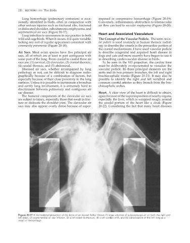
Lung hemorrhage (pulmonary contusion) is occa- imposed or compressive hemorrhage (Figure 20-19).
sionally identified in birds, often in conjunction with Conversely, inflammatory obstruction to intersaccular
other serious injuries such as fractured ribs, fractured air fl ow can lead to saccular emphysema (Figure 20-20).
or dislocated shoulder, subcutaneous emphysema, and
asymmetrical air sacs (Figure 20-17). Heart and Associated Vasculature
Lung infection is uncommon in our practice in both
wild and cage birds. When it occurs, it is quite variable, The Concept of the Vascular Pedicle. The term vascu-
lacking any sort of regular appearance consistent with lar pedicle is used routinely in human thoracic radiol-
community pneumonia (Figure 20-18). ogy to describe the vessels in the precardiac portion of
the cranial mediastinum. I have used vascular pedicle
Air Sacs. Most avian species have five principal air to describe congenital and acquired heart disease in
sacs, all of which are at least in part contiguous with dogs and cats and more recently have begun to use it
some part of the lung. From cranial to caudal these air in describing cardiovascular disease in birds.
sacs are: (1) cervical, (2) clavicular, (3) cranial thoracic, To be seen in the VD projection, the cardiac base
(4) caudal thoracic, and (5) abdominal. must be deliberately overpenetrated to visualize the
Diseased air sacs, whether accompanied by lung vascular pedicle. Its three principal elements are the
pathology or not, can be difficult to diagnose radio- aorta and its two primary branches, the left and right
graphically because of a combination of factors, but brachiocephalic trunks (Figure 20-21). It may also be
especially because of their close proximity to the lung possible to identify the right and left vertebral and
surfaces. Unless it is possible to incriminate a bronchus common carotid arteries as they branch from the bra-
and certify lung involvement, it is extremely hard to chiocephalic arches.
discriminate between pulmonary and contiguous air
sac disease. Heart. A clear view of the heart is difficult to obtain,
The humeral components of the clavicular air sacs again because of the superimposition of nearby organs,
are subject to injury, especially those that result in frac- especially the liver, which is wrapped snugly around
ture or dislocate the shoulder joint. The clavicular air the caudal portion of the heart like a cloak (Figure
sacs may also appear overly dense because of super- 20-22). Considering the fact that many heart diseases
Figure 20-17 • Ventrodorsal projection of the torso of an injured flicker shows (1) large volumes of subcutaneous air on both the right and
left sides, (2) asymmetrical air sac inflation, (3) a left-sided rib fracture, (4) a left cardiac shift, and (5) consolidation of the left lung as a
result of hemorrhage.
2/11/2008 11:08:51 AM
ch020-A02527.indd 220
ch020-A02527.indd 220 2/11/2008 11:08:51 AM