
Page 228 - Veterinary diagnostic imaging birds exotic pets wildlife
P. 228
224 SECTION I III The Birds
mass, although not all of these organs are always
visible in a particular radiograph (Figure 20-26).
Using Nature’s Contrast. Identification of the crop is
made easier when it contains a recent meal, particu-
larly if bones are present, for example, a mouse in the
gullet of a Merlin (Figure 20-27). The same is true of
the ventriculus, although in this location there is
usually more disintegration of whatever the bird has
eaten (Figure 20-28). High-density material, such as
bone, can also be seen in the bowel mass, but because
of ongoing digestion, it may be harder to specifi cally
identify. Cage birds and some wild birds may have grit
in their stomachs or bowel; grit is characterized radio-
graphically by its granular appearance and high
density (Figure 20-29)
Gastrointestinal Foreign Material. Of all the things a
wild bird might consume, lead is of the greatest concern
because of its toxicity. As a heavy metal, even small
quantities of lead are readily identifiable on a radio-
graph (Figure 20-30), while most pesticides, equally
harmful in their own right, are radiographically
invisible.
Assessment of the Digestive Tract with Barium and
Other Diagnostic Opaque Materials. Barium solu-
tions, or pastes, are the diagnostic opaque material of
choice for evaluating the alimentary canal. If a perfora-
tion is suspected, however, a nonionic organic iodine
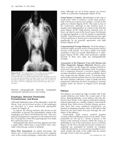
Figure 20-23 • The appearance of the central visceral pedicle in solution should be employed on the possibility that it
birds is highly species-dependent as exemplified in this may escape into the celomic cavity. A nonionic diag-
ventrodorsal view of a healthy great blue heron. Because the liver
is more caudally situated in this bird, more of the caudal aspect of nostic iodine solution is better than an ionic one because
the heart is apparent, especially the cardiac apex. For its part, the the ionic solution will draw water to itself and thus
liver forms more of a silhouette with the bowel mass than with temporarily dehydrate the bird. Examples of a limited
the heart. barium series are shown in Figures 20-31 through
20-33.
detected radiographically. Likewise, sonographic Kidneys
assessments typically prove unrevealing.
The kidneys are tucked up high on either side of the
caudal abdomen just below the cranial part of the
Esophagus, Stomach (Ventriculus-
Proventriculus), and Bowel pelvis. Radiographically, avian kidneys lack the dis-
tinctive bean shape of the typical mammalian kidney,
Although legitimate parts of the alimentary canal, the instead appearing as a variably shaped, somewhat ill-
throat, crop, and proximal portion of the esophagus defined mass. Some forms of kidney disease, particu-
are discussed in a more anatomically appropriate larly those that cause significant enlargement, can
context in Chapter 19. present as lameness because some of the nerves that
In raptors, the oblong stomach, also known as the innervate the hind limbs first pass through the sub-
ventriculus, or gizzard, lies immediately caudal to the stance of the kidneys.
esophagus, just above and behind the liver. This ana- Occasionally, birds receive nonfatal gunshot
tomical arrangement is apparent in the lateral but not wounds; pellet and BB guns seem to be favorites. These
the VD projection where the superimposed liver hides projectiles can end up in a variety of places: in the
the ventriculus from view. lumen of the stomach or bowel, within the cloaca, or
Radiographically, the bowel often appears more like embedded in one of the solid organs such as a kidney
a single object rather than a long hollow tube, espe- (Figure 20-34). Even though the precise location of a
cially when the gas content is minimal. projectile can sometimes be located, for example,
within the left kidney, it is not always possible to accu-
Plain Film Assessment. As stated previously, the rately predict the path of the pellet and the tissues
lateral view of the torso provides the most complete through which it may have passed.
view of the caudal esophagus, ventriculus, and bowel Text continued on p. 234.
2/11/2008 11:08:54 AM
ch020-A02527.indd 224
ch020-A02527.indd 224 2/11/2008 11:08:54 AM