Page 227 - Veterinary diagnostic imaging birds exotic pets wildlife
P. 227
CHAPTER 20  III The Torso  223


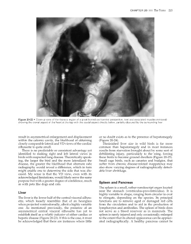
                    Figure 20-22  •  Close-up view of the thoracic region of a great horned owl (ventral perspective, keel and associated muscles removed)
                    showing the cranial aspect of the heart at the top with the caudal aspect directly below, partially obscured by the surrounding liver.


                    result in asymmetrical enlargement and displacement   or no doubt exists as to the presence of hepatomegaly
                    within the celomic cavity, the likelihood of obtaining   (Figure 20-24).
                    closely comparable lateral and VD views of the cardiac   Diminished liver size in wild birds is far more
                    silhouette is quite small.                           common than hepatomegaly and in most instances
                       There is no predictable or consistent advantage yet   results from starvation brought about by some sort of
                    identified to making right and left lateral views in   debilitating injury, particularly to the wing, forcing

                    birds with suspected lung disease. Theoretically speak-  these birds to become ground dwellers (Figure 20-25).
                    ing, the larger the bird and the more lateralized the   Small cage birds, such as canaries and budgies, that
                    disease, the greater the likelihood that alternate side   suffer from chronic disease-related inappetence may
                    radiography would reveal a difference, which in turn   also show varying degrees of radiographically detect-
                    might enable one to determine the side that was dis-  able liver shrinkage.
                    eased. My sense is that the VD view, even with its
                    acknowledged limitations, would likely serve the same
                    purpose but with a greater degree of confi dence, much   Spleen and Pancreas
                    as with pets like dogs and cats.
                                                                         The spleen is a small, rather nondescript organ located
                                                                         near the stomach (ventriculus-proventriculus). It is
                    Liver
                                                                         highly variable in shape, ranging from circular to oval
                    The liver is the lower half of the central visceral silhou-  to elongate, depending on the species. Its principal
                    ette, which loosely resembles that of an hourglass   functions are to remove aged or damaged red cells
                    when projected ventrodorsally, albeit a highly variable   from the circulation and to aid in the production of
                    one. As mentioned previously, the presence of an     lymphocytes and antibodies. The spleen of birds does
                    asymmetrical central visceral silhouette has yet to   not serve as a blood reservoir as in mammals. The
                    establish itself as a reliable indicator of either cardiac or   spleen is rarely injured and only occasionally enlarged
                    hepatic disease (Figure 20-23). If this is the case, it must   to the extent that its altered appearance can be appreci-
                    be acknowledged that there are instances where little   ated radiographically. A healthy pancreas cannot be


                                                                                                                      2/11/2008   11:08:53 AM
           ch020-A02527.indd   223                                                                                    2/11/2008   11:08:53 AM
           ch020-A02527.indd   223

