
Page 226 - Veterinary diagnostic imaging birds exotic pets wildlife
P. 226
222 SECTION I III The Birds
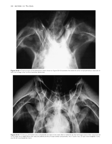
Figure 20-20 • Close-up view of the pneumonic raven shown in Figure 20-18 illustrates the extent to which emphysematous clavicular air
sacs can enlarge when outflow becomes obstructed.
Figure 20-21 • Close-up, penetrated view (a deliberate increase in kVp over what is called for by the technique chart) of the cranial portion
of the heart of an eagle shows the vascular pedicle and its principal arterial components: the (1) aortic root, (2) right brachiocephalic trunk,
and (3) left brachiocephalic trunk.
2/11/2008 11:08:52 AM
ch020-A02527.indd 222 2/11/2008 11:08:52 AM
ch020-A02527.indd 222