
Page 233 - Veterinary diagnostic imaging birds exotic pets wildlife
P. 233
CHAPTER 20 III The Torso 229
A
B
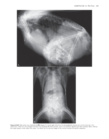
Figure 20-28 • A, Lateral and ventrodorsal (B) images of a great gray owl show the disintegrating carcass of a small mammal in the
ventriculus. Note how the enlarged stomach has displaced the liver cranially, and in the process, spread apart the individual lobes, making
the organ appear much wider than usual. The effect on the size and shape of the central visceral silhouette is dramatic.
2/11/2008 11:09:00 AM
ch020-A02527.indd 229 2/11/2008 11:09:00 AM
ch020-A02527.indd 229