Page 261 - Veterinary diagnostic imaging birds exotic pets wildlife
P. 261

CHAPTER 21  III Guinea Pigs  257


                                                A


                                          B


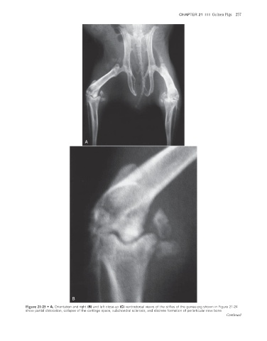
                    Figure 21-25  •  A, Orientation and right (B) and left close-up (C) ventrodorsal views of the stifles of the guinea pig shown in Figure 21-24
                    show partial dislocation, collapse of the cartilage space, subchondral sclerosis, and discrete formation of periarticular new bone.
                                                                                                                  Continued


                                                                                                                      2/11/2008   11:09:57 AM
           ch021-A02527.indd   257
           ch021-A02527.indd   257                                                                                    2/11/2008   11:09:57 AM
   256   257   258   259   260   261   262   263   264   265   266