
Page 353 - Veterinary diagnostic imaging birds exotic pets wildlife
P. 353
CHAPTER 30 III Acreage Pets 349
premature, immature, or “dysmature.” In the latter
instance, I never cease to be amazed at how often
normal carpal bones are mistakenly diagnosed as hypo-
plastic simply because the viewer is unfamiliar with
the projectional variations that typically accompany
limb curvatures.
Occasionally, limb curvature is seen, in which the
affected bones exhibit the characteristic changes of
rickets: flared metaphyses and abnormally widened
and irregular growth plates, which typically are found
throughout the appendicular skeleton (Figure 30-8).
Unlike the simple valgus and varus curvatures seen
with asymmetrical physeal growth, the rickets-like
cases show more complex deformities that often
include axial angulation. Unlike simple carpal (or com-
bined carpal-fetlock) curvatures, other important
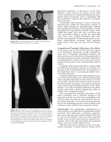
Figure 30-6 • A young alpaca and its student handlers pause
before entering the small animal x-ray suite. joints, such as the hips, are also involved (Figure
30-9).
Congenital and Traumatic Dislocation of the Elbow.
As crias grow, one or more of their joints may appear
to be developing abnormally, usually indicated by
some sort of nonpainful mechanical lameness that
often involves one or both elbows. Radiographically, it
is usually apparent the joint is abnormal, especially if
the opposite limb is normal and can be used for com-
parison, but the precise cause of the deformity may be
unclear.
Congenital dislocations of the elbow usually exhibit
the following radiographic features: (1) complete dis-
location of the humeroradial joint, (2) hypoplasia of
the radial head, and (3) a pronounced cranial curva-
ture of the olecranon.
Traumatic dislocation is also characterized by luxa-
tion of the radial head but can usually be distinguished
from congenital dislocation by the presence of a false
joint formed with the distal humeral shaft (as seen in
the flexed lateral projection) and marked hypoplasia
of the distal humeral epiphysis (Figure 30-10). Addi-
tionally, a cria with a traumatic dislocation of its elbow
usually cannot fully extend its injured leg, unlike an
animal with a congenital luxation.
Both congenital and traumatic dislocations of the
elbow eventually lead to dramatic changes in the
appearance of the involved bones, particularly their
cortices, which may become asymmetrically thick on
the load-bearing side. In the proximal ulna, participa-
tion in the remodeling process is most evident in the
semilunar notch, which becomes intensely sclerotic.
Osteomyelitis. The radiographic manifestations of
Figure 30-7 • Frontal view of the midforelimbs of an immature
alpaca centered on an imaginary perpendicular line drawn through osteomyelitis are no different in Camelids than in other
the center of the pectoral muscles. In this view, standardized to mammals: bone destruction, cavitations, sequestra-
the midsagittal line of the animal’s trunk, it is not only possible to tion, and a typically unsuccessful attempt by the host
determine what degree of angular deformity exists (varus/valgus) to wall off the infection with new bone. Sequestra
but as important, to determine whether there is also axial or
torsional deformity. This is termed the Nancy view in recognition usually occur in bones surrounded by minimal mus-
of the radiology technician who assisted in its development and culature, such as the large metacarpal and metatarsal
subsequent validation. bones, as occurs in horses (Figure 30-11).
Infections also occur commonly in the lower jaw,
usually secondary to dental infection (Figure 30-12)
2/11/2008 11:23:36 AM
ch030-A02527.indd 349 2/11/2008 11:23:36 AM
ch030-A02527.indd 349