
Page 377 - Veterinary diagnostic imaging birds exotic pets wildlife
P. 377
374 SECTION III III The Reptiles
Figure 32-10 • A Mexican black king snake lies coiled on an x-ray cassette awaiting radiography. Note the deformity in the center of the
lowermost coil, the result of a previous spinal fracture.
A
B
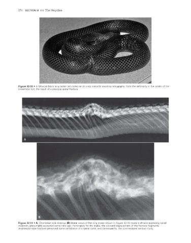
Figure 32-11 • A, Orientation and close-up (B) lateral views of the king snake shown in Figure 32-10 reveal a chronic-appearing spinal
malunion, presumably sustained some time ago. Fortunately for the snake, the outward displacement of the fracture fragments
(expression-type fracture) preserved some semblance of a spinal canal, and consequently, the cord escaped serious injury.
2/11/2008 11:26:19 AM
ch032-A02527.indd 374 2/11/2008 11:26:19 AM
ch032-A02527.indd 374