
Page 373 - Veterinary diagnostic imaging birds exotic pets wildlife
P. 373
370 SECTION III III The Reptiles
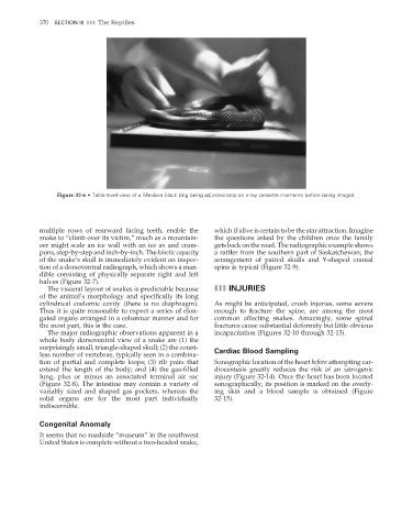
Figure 32-6 • Table-level view of a Mexican black king being adjusted atop an x-ray cassette moments before being imaged.
multiple rows of rearward facing teeth, enable the which if alive is certain to be the star attraction. Imagine
snake to “climb over its victim,” much as a mountain- the questions asked by the children once the family
eer might scale an ice wall with an ice ax and cram- gets back on the road. The radiographic example shows
pons, step-by-step and inch-by-inch. The kinetic capacity a rattler from the southern part of Saskatchewan; the
of the snake’s skull is immediately evident on inspec- arrangement of paired skulls and Y-shaped cranial
tion of a dorsoventral radiograph, which shows a man- spine is typical (Figure 32-9).
dible consisting of physically separate right and left
halves (Figure 32-7).
The visceral layout of snakes is predictable because III INJURIES
of the animal’s morphology and specifically its long
cylindrical coelomic cavity (there is no diaphragm). As might be anticipated, crush injuries, some severe
Thus it is quite reasonable to expect a series of elon- enough to fracture the spine, are among the most
gated organs arranged in a columnar manner and for common affecting snakes. Amazingly, some spinal
the most part, this is the case. fractures cause substantial deformity but little obvious
The major radiographic observations apparent in a incapacitation (Figures 32-10 through 32-13).
whole body dorsoventral view of a snake are (1) the
surprisingly small, triangle-shaped skull; (2) the count- Cardiac Blood Sampling
less number of vertebrae, typically seen in a combina-
tion of partial and complete loops; (3) rib pairs that Sonographic location of the heart before attempting car-
extend the length of the body; and (4) the gas-fi lled diocentesis greatly reduces the risk of an iatrogenic
lung, plus or minus an associated terminal air sac injury (Figure 32-14). Once the heart has been located
(Figure 32-8). The intestine may contain a variety of sonographically, its position is marked on the overly-
variably sized and shaped gas pockets, whereas the ing skin and a blood sample is obtained (Figure
solid organs are for the most part individually 32-15).
indiscernible.
Congenital Anomaly
It seems that no roadside “museum” in the southwest
United States is complete without a two-headed snake,
2/11/2008 11:26:16 AM
ch032-A02527.indd 370 2/11/2008 11:26:16 AM
ch032-A02527.indd 370