
Page 386 - Veterinary diagnostic imaging birds exotic pets wildlife
P. 386
CHAPTER 33 III Lizards 383
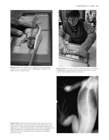
Figure 33-11 • The lizard shown in Figure 33-10 is secured and
settled on an x-ray cassette with a combination of gentle hand Figure 33-12 • One way to obtain a lateral projection of a lizard is
pressure and a wooden spoon. to simply place the animal on its side and then restrain in that
position long enough to make an exposure.
Figure 33-13 • Flexed dorsoventral view of the right femur of an
iguana suspected of having a fracture. Note that although the right
hind limb is not optimally positioned, it is adequately exposed, as
indicated by a readily distinguishable femoral cortex and medulla. This
desired appearance was achieved by reducing the radiographic
technique over what would normally be required to adequately
expose the torso.
2/11/2008 11:26:47 AM
ch033-A02527.indd 383 2/11/2008 11:26:47 AM
ch033-A02527.indd 383