
Page 390 - Veterinary diagnostic imaging birds exotic pets wildlife
P. 390
CHAPTER 33 III Lizards 387
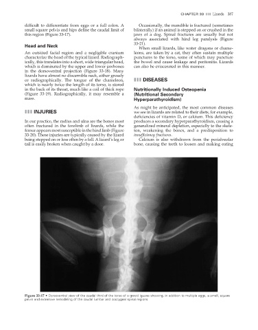
difficult to differentiate from eggs or a full colon. A Occasionally, the mandible is fractured (sometimes
small square pelvis and hips define the caudal limit of bilaterally) if an animal is stepped on or crushed in the
this region (Figure 33-17). jaws of a dog. Spinal fractures are usually but not
always associated with hind leg paralysis (Figure
33-21).
Head and Neck
When small lizards, like water dragons or chame-
An outsized facial region and a negligible cranium leons, are taken by a cat, they often sustain multiple
characterize the head of the typical lizard. Radiograph- punctures to the torso, some of which may puncture
ically, this translates into a short, wide triangular head, the bowel and cause leakage and peritonitis. Lizards
which is dominated by the upper and lower jawbones can also be eviscerated in this manner.
in the dorsoventral projection (Figure 33-18). Many
lizards have almost no discernible neck, either grossly
or radiographically. The tongue of the chameleon, III DISEASES
which is nearly twice the length of its torso, is stored
in the back of its throat, much like a coil of thick rope Nutritionally Induced Osteopenia
(Figure 33-19). Radiographically, it may resemble a (Nutritional Secondary
mass. Hyperparathyroidism)
As might be anticipated, the most common diseases
III INJURIES we see in lizards are related to their diets, for example,
deficiencies of vitamin D 3 or calcium. This defi ciency
In our practice, the radius and ulna are the bones most produces a secondary hyperparathyroidism, causing a
often fractured in the forelimb of lizards, while the generalized mineral depletion, especially to the skele-
femur appears most susceptible in the hind limb (Figure ton, weakening the bones, and a predisposition to
33-20). These injuries are typically caused by the lizard insuffi ciency fractures.
being stepped on or less often by a fall. A lizard’s leg or Calcium is also withdrawn from the perialveolar
tail is easily broken when caught by a door. bone, causing the teeth to loosen and making eating
Figure 33-17 • Dorsoventral view of the caudal third of the torso of a gravid iguana showing, in addition to multiple eggs, a small, square
pelvis and extensive remodeling of the caudal lumbar and coccygeal spinal regions.
2/11/2008 11:26:51 AM
ch033-A02527.indd 387 2/11/2008 11:26:51 AM
ch033-A02527.indd 387