
Page 395 - Veterinary diagnostic imaging birds exotic pets wildlife
P. 395
392 SECTION III III The Reptiles
A B
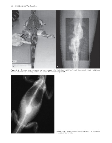
Figure 33-23 • A, Iguana, seen from above rear, has an angular deformity in the cranial third of its tail, the result of multiple insuffi ciency
fractures sustained over a year ago, as seen in a current dorsoventral radiograph (B).
Figure 33-24 • Near full-length dorsoventral view of an iguana with
a midintestinal enterolith.
2/11/2008 11:26:56 AM
ch033-A02527.indd 392 2/11/2008 11:26:56 AM
ch033-A02527.indd 392