
Page 398 - Veterinary diagnostic imaging birds exotic pets wildlife
P. 398
CHAPTER 33 III Lizards 395
A
C
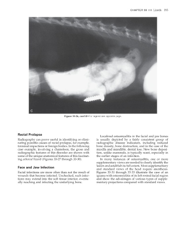
Figure 33-26, cont’d • For legend see opposite page.
Rectal Prolapse
Localized osteomyelitis in the facial and jaw bones
Radiography can prove useful in identifying or elimi- is usually depicted by a fairly consistent group of
nating possible causes of rectal prolapse, for example, radiographic disease indicators, including reduced
terminal impactions or foreign bodies. In the following bone density, bone destruction, and in the case of the
case example, involving a chameleon, the gross and maxilla and mandible, dental loss. New bone deposi-
radiographic features of this disorder are shown with tion, unlike mammals, is typically scant, especially in
some of the unique anatomical features of this fascinat- the earlier stages of an infection.
ing arboreal lizard (Figures 33-27 through 33-30). In many instances of osteomyelitis, one or more
supplementary views are needed to clearly identify the
lesion and establish its full extent. Most supplementary
Face and Jaw Infection
and standard views of the head require anesthesia.
Facial infections are more often than not the result of Figures 33-31 through 33-33 illustrate the case of an
wounds that become infected. Unchecked, such infec- iguana with osteomyelitis of its left rostral facial region
tions may extend into the soft tissue interior, eventu- and show the advantages of various types of supple-
ally reaching and infecting the underlying bone. mentary projections compared with standard views.
2/11/2008 11:27:00 AM
ch033-A02527.indd 395 2/11/2008 11:27:00 AM
ch033-A02527.indd 395