
Page 72 - Veterinary diagnostic imaging birds exotic pets wildlife
P. 72
68 SECTION I III The Birds
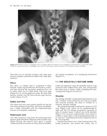
Figure 6-5 • Shoulder joints of a nestling snowy owl appear deficient because of the cartilaginous composition of its proximal humeri,
which are currently invisible on radiograph. Also note the characteristic diamond shape of the immature cervical vertebrae.
Thus there are no articular surfaces, only large gaps and appear incomplete, as if undergoing destruction
between partially ossified bones filled with vague gray (Figure 6-7).
shadows.
III THE SKELETALLY MATURE WING
Elbow
The elbow, or cubital, joint is comprised of three Unlike the immature wing, the skeletally mature wing
blunted, widely separated bones: the humerus, radius, possesses fully ossified bone ends, and consequently
and ulna. Because the associated epiphyses have yet the entire bone is clearly visible, including the inter-
to ossify, there are no visible joints, only hazy gaps vening joints (Figures 6-8 and 6-9).
(Figure 6-6, A). As the epiphyses ossify, the bone ends
become rounder and the intra-articular gaps become Shoulder
smaller (Figure 6-6, B).
The oblong, laterally compressed humeral head is set
into a shallow socket formed by the scapula caudally
Radius and Ulna
and coracoid cranially; the latter is overlain by a
The radius and ulna curve gently distally but like the common articular cartilage.
rest of the immature long bones, fall short of establish- The standard shoulder examination consists of a VD
ing a visible articulation with the carpometacarpus view (Figure 6-10), which can be supplemented by
because of incomplete ossifi cation. right and left VD oblique projections, especially in
cases of complex injury. A lateral view of the shoulder
(Figure 6-11) can prove indispensable when trying to
Radiocarpal Joint
confirm or deny injury to the scapulohumeral joint, or
Like other immature wing joints, the radiocarpal joint to “uncover” superimposed fracture fragments seen in
appears quite abnormal compared with its adult coun- the VD projection.
terpart because the apparent joint space is greatly Perhaps the clearest view of the humeral joint is the
widened and the bone ends are faint, almost cloudlike, frontal projection, also termed the leading edge or
2/11/2008 11:43:21 AM
ch006-A02527.indd 68 2/11/2008 11:43:21 AM
ch006-A02527.indd 68