
Page 75 - Veterinary diagnostic imaging birds exotic pets wildlife
P. 75
CHAPTER 6 III The Wing: Radiography and Normal Radiographic Anatomy 71
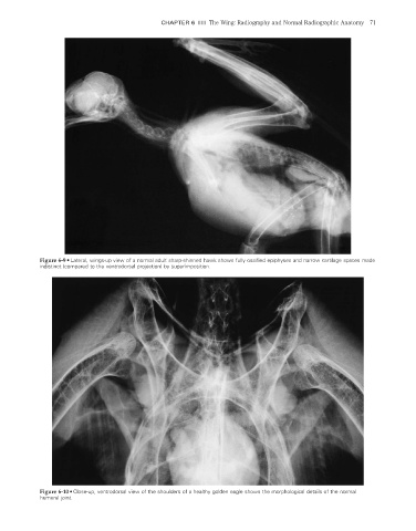
Figure 6-9 • Lateral, wings-up view of a normal adult sharp-shinned hawk shows fully ossified epiphyses and narrow cartilage spaces made
indistinct (compared to the ventrodorsal projection) by superimposition.
Figure 6-10 • Close-up, ventrodorsal view of the shoulders of a healthy golden eagle shows the morphological details of the normal
humeral joint.
2/11/2008 11:43:24 AM
ch006-A02527.indd 71 2/11/2008 11:43:24 AM
ch006-A02527.indd 71