
Page 79 - Veterinary diagnostic imaging birds exotic pets wildlife
P. 79
CHAPTER 6 III The Wing: Radiography and Normal Radiographic Anatomy 75
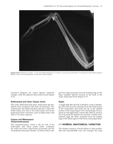
Figure 6-15 • Projectional variation. In this ventrodorsal projection of a hawk’s wing, the proximal half of the radial and ulnar bodies appears
bowed, while the distal diaphyses of both bones seem straight.
increased obliquity, the radius appears relatively gial first digit protrudes from the leading edge of the
straight, while the adjacent ulna looks bowed (Figure large irregular tubercle located on the head of the
6-16). major metacarpal bone (Figure 6-17).
Radiocarpal and Ulnar Carpal Joints Digits
The avian radiocarpal and ulnar carpal joints are sim- A single digit (the second), formed by a pair of phalan-
plified when compared with those of mammals. Two ges (fi rst and second), emanates from the distal aspect
carpal bones, the radial and ulnar, directly connect the of the metacarpus and forms the tip of the skeletal
distal radius and ulna to the fused major and minor portion of the wing (Figure 6-18). Interestingly, the ter-
metacarpal bones and form a pair of hinge joints with minal second digit barely reaches the midway mark of
little or no rotary capacity. the wing when the feathers are included. Another rudi-
mentary digit, the third, originates from the trailing
edge of the distal aspect of the minor metacarpal bone.
Carpus and Metacarpus
(Carpometacarpus)
The carpometacarpus, which is the far side of the III NORMAL ANATOMICAL VARIATION
radiocarpal and ulnar carpal joints, comprises
the radial and ulnar carpal bones proximally and the The skeletal anatomy of birds differs in both predict-
streamlined metacarpus distally. An abbreviated vesti- able and unpredictable ways. For example: the wing
2/11/2008 11:43:28 AM
ch006-A02527.indd 75 2/11/2008 11:43:28 AM
ch006-A02527.indd 75