
Page 136 - Withrow and MacEwen's Small Animal Clinical Oncology, 6th Edition
P. 136
CHAPTER 6 Diagnostic Imaging in Oncology 115
be used to predict malignancy, but this finding is not consis-
tent. 32,33 In one study of liver masses, arterial hypervascularity was
noted in the majority of hepatocellular carcinoma masses, but not
VetBooks.ir other benign or malignant hepatic masses. Multiphase evalua-
34
tion of enhancement patterns with contrast-enhanced ultrasound
can be used to differentiate between benign and malignant lesions.
In both the liver and spleen, a nodule that remains hypoechoic in
both the early vascular phase (5–10 seconds after injection) and
late vascular phase (25–30 seconds after injection) is more com-
monly seen with malignant lesions whereas benign nodules most
often become isoechoic to the surrounding parenchyma in both
phases. 21,32,33,35,36 However, this distinction is less clear for renal
37
lesions. This differential contrast pattern has also been shown to
increase the sensitivity for the diagnosis of liver metastasis. 31
Elastography uses ultrasound to assess the relative hardness of
tissues. This is an emerging technology in both human and vet-
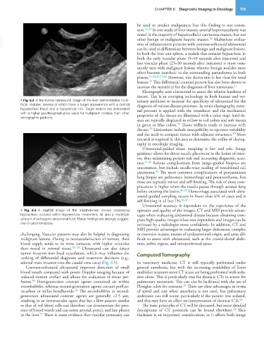
• Fig. 6.3 A transverse ultrasound image of the liver demonstrates multi- erinary medicine to increase the specificity of ultrasound for the
focal nodules, several of which have a target appearance with a central diagnosis of various disease processes. In strain elastography, exter-
hyperechoic focus and a hypoechoic rim. Target lesions are associated nal pressure is applied with the transducer and the mechanical
with a higher positive-predicative value for malignant nodules than other properties of the tissues are illustrated with a color map; hard tis-
sonographic patterns.
sues are typically displayed in yellow to red colors and soft tissues
in green to blue colors. Tissue stiffness tends to increase with
38
disease. Limitations include susceptibility to operator variability
39
40
and the need to compare tissues with adjacent structures. More
research is required in this area to determine the utility of elastog-
raphy in oncologic imaging.
Ultrasound-guided tissue sampling is fast and safe. Image
guidance allows for direct needle placement in the lesion of inter-
est, thus minimizing patient risk and increasing diagnostic accu-
racy. 41,42 Serious complications from image-guided biopsies are
uncommon, but include needle-tract seeding of transitional cell
carcinoma. The most common complications of percutaneous
43
lung biopsy are pulmonary hemorrhage and pneumothorax, but
these are typically minor and self-limiting. The risk of these com-
plications is higher when the needle passes through aerated lung
before entering the lesion. 44,45 Hemorrhage associated with ultra-
sound guided sampling occurs in fewer than 6% of cases and is
self-limiting in all but 1%. 46,47
Ultrasound accuracy is dependent on the experience of the
• Fig. 6.4 A sagittal image of the midabdomen shows coalescing operator and quality of the images. CT and MRI provide advan-
hypoechoic nodules within hyperechoic mesenteric fat and a moderate tages when evaluating abdominal disease because obtaining com-
amount of echogenic abdominal fluid. These findings are strongly sugges- plete high-quality images is less user dependent and images can be
tive of carcinomatosis. reviewed by a radiologist more confidently. In addition, CT and
MRI provide advantages in evaluating larger abdomens, complex
challenging. Vascular patterns may also be helpful in diagnosing or extensive masses, masses of undetermined origin, and areas dif-
malignant lesions. Owing to neovascularization of tumors, their ficult to assess with ultrasound, such as the cranial-dorsal abdo-
blood supply tends to be more tortuous, with higher velocities men, pelvic region, and retroperitoneal space.
than noted in normal tissue. 26–29 Ultrasound can also detect
tumor invasion into local vasculature, which may influence the Computed Tomography
ranking of differential diagnoses and treatment decisions (e.g.,
adrenal mass invasion into the caudal vena cava) (Fig. 6.5). In veterinary medicine, CT is still typically performed under
Contrast-enhanced ultrasound improves detection of small general anesthesia, but with the increasing availability of faster
blood vessels compared with power Doppler imaging because of multislice scanners more CT scans are being performed with seda-
reduced motion artifact and allows for evaluation of tissue per- tion alone. This is particularly true for thoracic CTs to screen for
fusion. First-generation contrast agents contained air within pulmonary metastasis. This can also be facilitated with the use of
30
microbubbles, whereas second-generation agents contain perfluo- Plexiglas tubes for restraint. There are clear advantages in terms
48
31
rocarbon or sulfur hexafluoride. The microbubbles in second- of speed and cost when anesthesia is not used, but pulmonary
generation ultrasound contrast agents are generally <2.5 μm, atelectasis can still occur, particularly if the patient was sedated,
resulting in an intravascular agent that has a flow pattern similar and this may have an effect on interpretation of thoracic CTs. 48
to that of red blood cells and therefore can demonstrate the pres- The basic principles of CT will be discussed, but more detailed
ence of blood vessels and can assess arterial, portal, and late phases descriptions of CT protocols can be found elsewhere. Slice
49
in the liver. There is some evidence that vascular tortuosity can thickness is an important consideration, as it affects both image
31