Page 180 - Withrow and MacEwen's Small Animal Clinical Oncology, 6th Edition
P. 180
CHAPTER 9  Biopsy and Sentinel Lymph Node Mapping Principles  159


                                                                 The stab incision can be sutured with a single interrupted suture.
                                                                 The tissue is gently removed from the instrument with a scalpel
                                                                 blade or hypodermic needle and placed in formalin and/or alter-
  VetBooks.ir                                                    native media (e.g., culture media) or snap frozen as necessary. For
                                                                 smaller-gauge needle-core biopsy instruments, the tissue may be
                                                                 flushed off the needle with saline. Samples may be gently rolled on
                                                                 a glass slide for cytologic preparations before fixation. With expe-
                                                                 rience, the operator can generally tell from the appearance of the
                                                                 core sample whether diagnostic material has been attained. Small,
                                                                 discontinuous segments of tissue and fluid within the trough will
                                                                 only rarely be diagnostic and usually imply the need for incisional
                                                                 biopsy. Soft tissue sarcomas in particular may not yield good tissue
                                                                 cores because of necrosis and fibrous septa that often permeate the
                                                                 mass. Cystic masses are also problematic.
                                                                   Needle biopsy  tracts are probably a minimal  risk for local
                                                                 tumor seeding but should be removed en bloc with the tumor at
                                                                 subsequent resection. Therefore it is important to plan where the
                                                                 stab incision and needle biopsy tract should be placed to make
                                                                 subsequent excision simpler. Avoid excessive tunneling through
                                                                 uninvolved tissues by choosing the most direct path from the skin
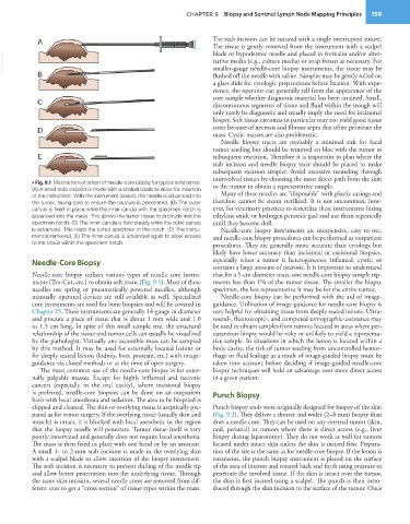
           • Fig. 9.1  Mechanism of action of needle-core biopsy for typical solid tumor.   to the tumor to obtain a representative sample.
           (A) A small stab incision is made with a scalpel blade to allow for insertion   Many of these needles are “disposable” with plastic casings and
           of the instrument. With the instrument closed, the needle is advanced into
           the tumor, taking care to ensure the capsule is penetrated. (B) The outer   therefore cannot be steam sterilized. It is not uncommon, how-
           canula is fixed in place while the inner canula with the specimen notch is   ever, for veterinary practices to resterilize these instruments (using
           advanced into the mass. This allows the tumor tissue to protrude into the   ethylene oxide or hydrogen peroxide gas) and use them repeatedly
           specimen notch. (C) The inner canula is held steady while the outer canula   until they become dull.
           is advanced. This traps the tumor specimen in the notch. (D) The instru-  Needle-core biopsy instruments are inexpensive, easy to use,
           ment is removed. (E) The inner canula is advanced again to allow access   and needle-core biopsy procedures can be performed as outpatient
           to the tissue within the specimen notch.              procedures. They are generally more accurate than cytology but
                                                                 likely have lower accuracy than incisional or excisional biopsies,
           Needle-Core Biopsy                                    especially when a tumor is heterogeneous, inflamed, cystic, or
                                                                 contains a large amount of necrosis. It is important to understand
           Needle-core biopsy utilizes various types of needle core instru-  that for a 5-cm diameter mass, one needle-core biopsy sample rep-
           ments (Tru-Cut, etc.) to obtain soft tissue (Fig. 9.1). Most of these   resents less than 1% of the tumor tissue. The smaller the biopsy
           needles use spring or pneumatically powered needles, although   specimen, the less representative it may be for the entire tumor.
           manually operated devices are still available as well. Specialized   Needle-core biopsy can be performed with the aid of image-
           core instruments are used for bone biopsies and will be covered in   guidance. Utilization of image-guidance for needle-core biopsy is
           Chapter 25. These instruments are generally 14-gauge in diameter   very helpful for obtaining tissue from deeply seated lesions. Ultra-
           and procure a piece of tissue that is about 1 mm wide and 1.0   sound-, fluoroscopic-, and computed tomographic-assistance may
           to 1.5 cm long. In spite of this small sample size, the structural   be used to obtain samples from tumors located in areas where per-
           relationship of the tissue and tumor cells can usually be visualized   cutaneous biopsy would be risky or unlikely to yield a representa-
           by the pathologist. Virtually any accessible mass can be sampled   tive sample. In situations in which the lesion is located within a
           by this method. It may be used for externally located lesions or   body cavity, the risk of tumor seeding from uncontrolled hemor-
           for deeply seated lesions (kidney, liver, prostate, etc.) with image-  rhage or fluid leakage as a result of image-guided biopsy must be
           guidance via closed methods or at the time of open surgery.  taken into account before deciding if image-guided needle-core
             The most common use of the needle-core biopsy is for exter-  biopsy techniques will hold an advantage over more direct access
           nally  palpable  masses.  Except  for highly  inflamed  and  necrotic   in a given patient.
           cancers (especially in the oral cavity), where incisional biopsy
           is preferred, needle-core biopsies can be done on an outpatient   Punch Biopsy
           basis with local anesthesia and sedation. The area to be biopsied is
           clipped and cleaned. The skin or overlying tissue is aseptically pre-  Punch biopsy tools were originally designed for biopsy of the skin
           pared as for minor surgery. If the overlying tissue (usually skin and   (Fig. 9.2). They deliver a shorter and wider (2–8 mm) biopsy than
           muscle) is intact, it is blocked with local anesthetic in the region   does a needle core. They can be used on any external tumor (skin,
           that the biopsy needle will penetrate. Tumor tissue itself is very   oral, perianal) or tumors where there is direct access (e.g., liver
           poorly innervated and generally does not require local anesthesia.   biopsy during laparotomy). They do not work as well for tumors
           The mass is then fixed in place with one hand or by an assistant.   located under intact skin unless the skin is incised first. Prepara-
           A small 1- to 2-mm stab incision is made in the overlying skin   tion of the site is the same as for needle-core biopsy. If the lesion is
           with a scalpel blade to allow insertion of the biopsy instrument.   cutaneous, the punch biopsy instrument is placed on the surface
           The stab incision is necessary to prevent dulling of the needle tip   of the area of interest and rotated back and forth using pressure to
           and allow better penetration into the underlying tissue. Through   penetrate the involved tissue. If the skin is intact over the tumor,
           the same skin incision, several needle cores are removed from dif-  the skin is first incised using a scalpel. The punch is then intro-
           ferent sites to get a “cross section” of tissue types within the mass.   duced through the skin incision to the surface of the tumor. Once

