Page 237 - Withrow and MacEwen's Small Animal Clinical Oncology, 6th Edition
P. 237
216   PART III    Therapeutic Modalities for the Cancer Patient


  VetBooks.ir


                   A                                           B
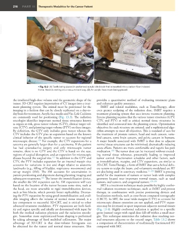
                          • Fig. 13.3  (A) Teeth being placed in preformed acrylic bite block that is inserted into a carbon fiber indexed
                          frame. Neck is resting on a vacuum-lock bag. (B) An acrylic face mask being placed.


         the irradiated high-dose volume and the geometric shape of the   provides a quantitative method of evaluating treatment plans
         tumor. 3D-CRT requires importation of CT images into a treat-  and enhances quality assurance.
         ment planning system. The animal must be positioned for the   IMRT and related modalities, such as TomoTherapy, allow
         imaging in a fashion that can be closely replicated on a day-to-  even greater sculpting of the radiation dose. IMRT requires a
         day basis for treatment. Acrylic face masks and Vac-Lok Cushions     treatment planning system that uses inverse treatment planning.
         are commonly used for positioning (Fig. 13.3). The radiation   Inverse planning requires that the various tumor structures (GTV,
         oncologist identifies important normal tissue structures known   CTV, and PTV) as well as critical normal tissue structures be
         as organs at risk, gross tumor volume (GTV), clinical target vol-  identified and contoured into the planning system. Optimization
         ume (CTV), and planning target volume (PTV) on these images.   objectives for each structure are entered, and a sophisticated algo-
         By definition, the GTV only includes gross tumor whereas the   rithm attempts to meet all objectives. This is standard of care for
         CTV includes the GTV plus an expansion based on the known   the treatment of prostate tumors, head and neck cancers, verte-
         clinical behavior of the specific tumor to account for regional   bral cancers, some brain cancers, and pelvic cancers in humans.
         microscopic disease. 121  For example, the CTV expansions for a   A major benefit associated with IMRT is that dose to adjacent
         sarcoma are generally larger than for a carcinoma. If the patient   normal tissue structures can be minimized, dramatically reducing
         has had cytoreductive surgery and only microscopic tumor   acute effects. Patients are more comfortable and require less pain
         remains, there is no GTV and the CTV is based on the scar,   medication. 122  The tumor dose can be increased without exceed-
         regions of surgical disruption, and an expansion for microscopic   ing normal tissue tolerance, presumably leading to improved
         disease beyond the surgical site. 121  In addition to the GTV and   tumor control. Fractionation schedules and other factors, such
         CTV, the PTV includes expansion for an internal margin that   as immobilization, margins, and CTV expansions, are similar to
         accounts for variations in size and shape relative to anatomic   3D-CRT. TomoTherapy, a form of IMRT that uses a helical deliv-
         landmarks (e.g., filling of bladder, respiratory movements) and   ery system to sculpt the beam, and volumetric-modulated arc RT
         set-up margin (SM). The SM accounts for uncertainties in   are also being used in veterinary medicine. 11,123  IMRT is proving
         patient positioning and alignment during planning imaging and   useful for the treatment of tumors or tumor beds with complex
         subsequent treatments. 121  The better the immobilization device,   geometry located near important normal tissues, such as nasal
         the smaller the SM expansion can be. SM expansions will vary   tumors, oral tumors, and urogenital tumors. 123,124
         based on the location of the tumor because some sites, such as   SRT is a treatment technique made possible by highly confor-
         the head, are more amenable to rigid immobilization devices,   mal radiation treatment techniques, such as IMRT and proton
         such as bite blocks, which provide better replicability. Decreas-  therapy, in combination with precise positioning devices and
         ing the PTV expansion by using good immobilization and avail-  localization methods, such as cone-beam computed tomography
         able imaging affects the volume of normal tissue treated, is a   (CBCT). In SRT, the usual wide margins (CTV) to account for
         key component to successful 3D-CRT, and is critical to other   microscopic disease extension are not applied, and PTV expan-
         advanced treatment modalities. PTV expansions are both facil-  sion may be decreased to spare important adjacent normal tissues
         ity and region specific and ideally are determined with input by   (Fig. 13.4). Highly conformal radiation is applied to a specific
         both the medical radiation physicist and the radiation oncolo-  gross (tumor) target with rapid dose fall-off within a small mar-
         gist. Somewhat more sophisticated beam shaping is performed   gin. This technique minimizes the radiation dose reaching nor-
         by taking advantage of fixed multileaf collimators. The major   mal structures adjacent to the treated target. Table 13.2 shows
         advantage  of  3D-CRT  is  that  dose-volume  histograms  can   a comparison of characteristics of traditionally fractionated RT
be obtained for the tumor and normal tissue structures. This compared with SRT. 

