
Page 378 - Withrow and MacEwen's Small Animal Clinical Oncology, 6th Edition
P. 378
356 PART IV Specific Malignancies in the Small Animal Patient
Institute of Pathology publication on the histologic classification
of skin tumors. 72
VetBooks.ir Basal Cell Carcinoma
The true incidence of BCC in both dogs and cats is unknown. The
different tumors previously categorized as BCTs are difficult to dis-
tinguish histologically and cytologically, both in determining the cat-
egory of tumor and in differentiating benign lesions from malignant.
On cytology BCTs can contain inflammatory cells, squamous cells,
sebaceous epithelial cells, melanin, and melanophages, and these cells
can express the criteria of malignancy. Well-differentiated fibroblasts,
reactive fibroblasts, and mast cells may also be present on cytologic
83
examination. The inability to distinguish the subtypes on cytology
has led to the suggestion that these tumors be called “cutaneous basi-
lar epithelial neoplasms” when they are evaluated by cytology alone. 84
In histopathologic evaluation, tumors sometimes can be
grossly differentiated based on growth pattern. The epithelial
85
membrane glycoprotein BerEP4 is highly specific for BCC versus
SCC or cystic-solid ductular tumors for the diagnosis of BCC in
people. Cytokeratin 8 (CAM5.2) is used in human tumors to
59
identify tumors with sweat gland epithelial differentiation; it also
has been used to differentiate a BCC from a solid-cystic ductular
tumor in a dog. However, validation of these IHC markers in a • Fig. 19.1 Fibropapilloma (sarcoid) on the face of a cat. (From Miller WH
84
larger veterinary population remains to be performed. Jr., Griffin CE, Campbell KL: Muller & Kirk’s small animal dermatology, ed
BCC is rare in dogs. In two studies reporting BCC or basal cell 7, St Louis, 2013, Saunders.)
epithelioma (benign variant), the incidence ranged from 6% to
8% of all skin tumors; however, it is unclear whether trichoblas- Pulmonary metastasis has been documented in one cat with a
89
tomas were included in these studies. 10,11 Breeds reported to be at BCC. Nucleomorphometric analysis was able to predict local
90
increased risk for BCC included cocker spaniel and poodle in one tumor recurrence in one study of 24 cats with BCC. Overall, the
study, and spaniels were overrepresented in another study; how- likelihood of metastasis with BCC in cats appears low.
ever, no breed predispositions were identified in other studies. 9,10 Treatment for BCC is wide surgical excision, which often results
Clinically, these tumors present as plaques or nodules, often in long-term control. In people with masses less than 2 cm, surgi-
darkly pigmented. The overlying skin may be alopecic and intact cal margins of 4 mm result in a high cure rate. However, surgical
or ulcerated. In dogs the median age is 9 years; however, dogs of all margins of 5 to 10 mm are recommended in cats and dogs, when
ages may be affected. 10,11 The three recognized histologic subtypes possible, because intraoperative margin assessment cannot be per-
91
are solid, keratinizing, and clear cell. 85 formed. The data is limited, but adjuvant radiation therapy (RT)
In dogs BCC is considered a low-grade malignancy. Although and doxorubicin have been used to treat BCCs in two cats; although
local recurrence of this tumor after surgical excision has been their contribution to tumor control and survival are unknown.
89
reported, there are no reports of metastasis. Morphometric analy-
sis of cell nuclei has been reported to be useful in differentiating Basosquamous Cell Carcinoma
BCC with the potential for local recurrence from BCC that is Histologically basosquamous cell carcinoma has characteristics of
unlikely to recur. 86 both SCC and BCC. Clinically it is indistinguishable from both
In cats BCCs are now thought to be rare, and many feline BCTs BCC and SCC. The true incidence and clinical behavior of this
are being reclassified as either solid-cystic apocrine ductular adenomas tumor in dogs and cats are unknown.
87
(approximately 60%) or trichoblastomas (approximately 40%).
However, given the preponderance of the literature referring to BCTs Papillomas
in cats, this group of tumors is discussed here with the realization
that the population is actually not homogenous. These tumors com- Papillomas are benign epidermal proliferative lesions that often
prise some of the most common solid tumors in cats, second only to are associated with PPV infection. They are considered rare in
34
2
mammary tumors in one large study. They represent 10% to 26% the cat and dog. Papillomas typically have an exophytic growth
92
of feline skin tumors. 2,6,9,88 They are reported in middle-aged cats pattern. Another benign variant is the inverted papilloma, which
with a mean age of 9.6 to 10.8 years. 2,6,88 One study reported a pre- grows into the subcutaneous tissue rather than externally. Cuta-
2
disposition to BCT in Siamese cats, and a second study showed an neous papillomas are typically found in younger dogs with an
38
increased number of these tumors in long-haired cats (Himalayans, average age of 3.2 years. Surgical excision is usually curative,
domestic long-hair, and Persian cats) ; however, a third study did but some of these lesions spontaneously regress. In patients with
93
88
not find a breed predisposition. BCC can appear anywhere on the multiple lesions, azithromycin has been shown to be effective. 94
6
body but may have a predilection for the head and neck. 6,88 BCC can In cats a particular type of papilloma, the fibropapilloma, is
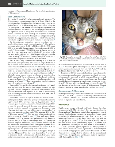
appear pigmented and may clinically resemble melanoma. seen. These tumors demonstrate a proliferation of mesenchymal
44
Clinically most tumors classified as BCTs appear benign in cells covered by hyperplastic epithelium. Evaluation for PPV
behavior. Malignant BCTs have been described in 10 of 97 cats demonstrated a nonproductive infection of the mesenchymal
95
in one series based on the presence of stromal invasion, vascu- cells. Feline fibropapillomas may be more similar to equine sar-
2
lar invasion, necrosis, a high mitotic index, and LN metastasis. coids than papillomas (Fig. 19.1). 