
Page 1085 - Veterinary Immunology, 10th Edition
P. 1085
VetBooks.ir
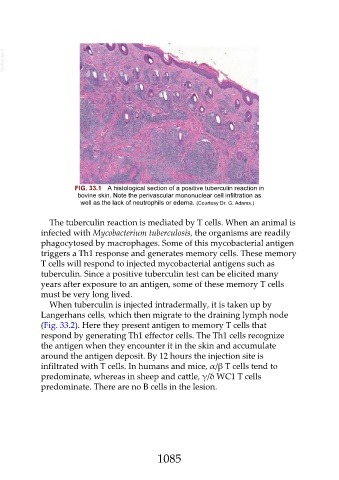
FIG. 33.1 A histological section of a positive tuberculin reaction in
bovine skin. Note the perivascular mononuclear cell infiltration as
well as the lack of neutrophils or edema. (Courtesy Dr. G. Adams.)
The tuberculin reaction is mediated by T cells. When an animal is
infected with Mycobacterium tuberculosis, the organisms are readily
phagocytosed by macrophages. Some of this mycobacterial antigen
triggers a Th1 response and generates memory cells. These memory
T cells will respond to injected mycobacterial antigens such as
tuberculin. Since a positive tuberculin test can be elicited many
years after exposure to an antigen, some of these memory T cells
must be very long lived.
When tuberculin is injected intradermally, it is taken up by
Langerhans cells, which then migrate to the draining lymph node
(Fig. 33.2). Here they present antigen to memory T cells that
respond by generating Th1 effector cells. The Th1 cells recognize
the antigen when they encounter it in the skin and accumulate
around the antigen deposit. By 12 hours the injection site is
infiltrated with T cells. In humans and mice, α/β T cells tend to
predominate, whereas in sheep and cattle, γ/δ WC1 T cells
predominate. There are no B cells in the lesion.
1085