
Page 1086 - Veterinary Immunology, 10th Edition
P. 1086
VetBooks.ir
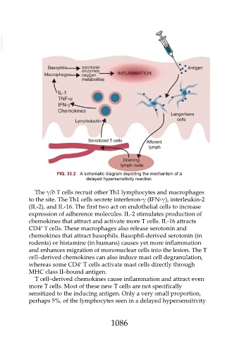
FIG. 33.2 A schematic diagram depicting the mechanism of a
delayed hypersensitivity reaction.
The γ/δ T cells recruit other Th1 lymphocytes and macrophages
to the site. The Th1 cells secrete interferon-γ (IFN-γ), interleukin-2
(IL-2), and IL-16. The first two act on endothelial cells to increase
expression of adherence molecules. IL-2 stimulates production of
chemokines that attract and activate more T cells. IL-16 attracts
+
CD4 T cells. These macrophages also release serotonin and
chemokines that attract basophils. Basophil-derived serotonin (in
rodents) or histamine (in humans) causes yet more inflammation
and enhances migration of mononuclear cells into the lesion. The T
cell–derived chemokines can also induce mast cell degranulation,
+
whereas some CD4 T cells activate mast cells directly through
MHC class II–bound antigen.
T cell–derived chemokines cause inflammation and attract even
more T cells. Most of these new T cells are not specifically
sensitized to the inducing antigen. Only a very small proportion,
perhaps 5%, of the lymphocytes seen in a delayed hypersensitivity
1086