
Page 1116 - Veterinary Immunology, 10th Edition
P. 1116
rejected, although the reverse is not the case. This is because male
VetBooks.ir cells carry an antigen coded for by genes on the Y chromosome,
called the H-Y antigen.
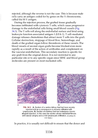
During the rejection process, the grafted tissue gradually
becomes infiltrated with cytotoxic T cells, which cause progressive
damage to the endothelial cells lining small blood vessels (Fig.
34.3). The T cells roll along the endothelial surface and bind using
leukocyte function-associated antigen-1 (LFA-1). T cell–mediated
damage releases chemokines that attract more T cells into the graft.
Cellular destruction, stoppage of blood flow, hemorrhage, and
death of the grafted organ follow thrombosis of these vessels. The
blood vessels of second organ grafts become blocked even more
rapidly as a result of the action of antibodies and complement on
the vascular endothelium. This secondary reaction is specific for
any graft from the original donor. It is not restricted to any
particular site or to any specific organ since MHC and blood group
molecules are present on most nucleated cells.
FIG. 34.3 A, Section of a canine kidney that had been acutely
rejected and as a consequence is densely infiltrated with
lymphocytes. B, Section of a kidney that has undergone chronic
allograft rejection. In this case the section shows interstitial fibrosis
with tubular atrophy and a mild lymphocytic infiltration. (Courtesy Dr.
A.E. Kyles.)
In practice, it is usually not difficult to ensure that the donor and
1116