
Page 161 - Veterinary Immunology, 10th Edition
P. 161
VetBooks.ir
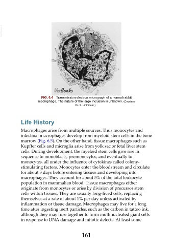
FIG. 6.4 Transmission electron micrograph of a normal rabbit
macrophage. The nature of the large inclusion is unknown. (Courtesy
Dr. S. Linthicum.)
Life History
Macrophages arise from multiple sources. Thus monocytes and
intestinal macrophages develop from myeloid stem cells in the bone
marrow (Fig. 6.5). On the other hand, tissue macrophages such as
Kupffer cells and microglia arise from yolk sac or fetal liver stem
cells. During development, the myeloid stem cells give rise in
sequence to monoblasts, promonocytes, and eventually to
monocytes, all under the influence of cytokines called colony-
stimulating factors. Monocytes enter the bloodstream and circulate
for about 3 days before entering tissues and developing into
macrophages. They account for about 5% of the total leukocyte
population in mammalian blood. Tissue macrophages either
originate from monocytes or arise by division of precursor stem
cells within tissues. They are usually long-lived cells, replacing
themselves at a rate of about 1% per day unless activated by
inflammation or tissue damage. Macrophages may live for a long
time after ingesting inert particles, such as the carbon in tattoo ink,
although they may fuse together to form multinucleated giant cells
in response to DNA damage and mitotic defects. At least some
161