
Page 336 - Veterinary Immunology, 10th Edition
P. 336
VetBooks.ir
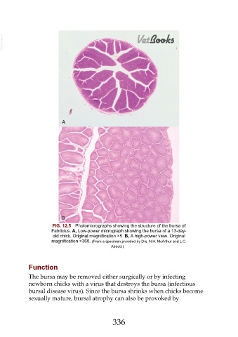
FIG. 12.5 Photomicrographs showing the structure of the bursa of
Fabricius. A, Low-power micrograph showing the bursa of a 13-day-
old chick. Original magnification ×5. B, A high-power view. Original
magnification ×360. (From a specimen provided by Drs. N.H. McArthur and L.C.
Abbott.)
Function
The bursa may be removed either surgically or by infecting
newborn chicks with a virus that destroys the bursa (infectious
bursal disease virus). Since the bursa shrinks when chicks become
sexually mature, bursal atrophy can also be provoked by
336