
Page 338 - Veterinary Immunology, 10th Edition
P. 338
VetBooks.ir
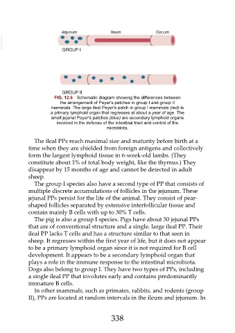
FIG. 12.6 Schematic diagram showing the differences between
the arrangement of Peyer's patches in group I and group II
mammals. The large ileal Peyer's patch in group I mammals (red) is
a primary lymphoid organ that regresses at about a year of age. The
small jejunal Peyer's patches (blue) are secondary lymphoid organs
involved in the defense of the intestinal tract and control of the
microbiota.
The ileal PPs reach maximal size and maturity before birth at a
time when they are shielded from foreign antigens and collectively
form the largest lymphoid tissue in 6-week-old lambs. (They
constitute about 1% of total body weight, like the thymus.) They
disappear by 15 months of age and cannot be detected in adult
sheep.
The group I species also have a second type of PP that consists of
multiple discrete accumulations of follicles in the jejunum. These
jejunal PPs persist for the life of the animal. They consist of pear-
shaped follicles separated by extensive interfollicular tissue and
contain mainly B cells with up to 30% T cells.
The pig is also a group I species. Pigs have about 30 jejunal PPs
that are of conventional structure and a single, large ileal PP. Their
ileal PP lacks T cells and has a structure similar to that seen in
sheep. It regresses within the first year of life, but it does not appear
to be a primary lymphoid organ since it is not required for B cell
development. It appears to be a secondary lymphoid organ that
plays a role in the immune response to the intestinal microbiota.
Dogs also belong to group I. They have two types of PPs, including
a single ileal PP that involutes early and contains predominantly
immature B cells.
In other mammals, such as primates, rabbits, and rodents (group
II), PPs are located at random intervals in the ileum and jejunum. In
338