
Page 346 - Veterinary Immunology, 10th Edition
P. 346
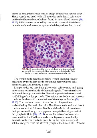
center of each paracortical cord is a high endothelial venule (HEV).
  VetBooks.ir  These vessels are lined with tall, rounded endothelial cells quite

               unlike the flattened endothelium found in other blood vessels (Fig.
               12.12). HEVs are surrounded by concentric layers of fibroblastic

               reticular cells and a narrow space called the perivenular channel.


                            FIG. 12.12  A section of human tonsil showing a high endothelial
                            venule with its characteristic high, rounded endothelial cells. Note
                                the lymphocytes emigrating between the endothelial cells.


                  The lymph node medulla contains lymph-draining sinuses

               separated by medullary cords containing many plasma cells,
               macrophages, and memory T cells.
                  Lymph nodes are very busy places with cells coming and going
               in response to a multitude of chemical signals. These signals are
               delivered through the reticular fibers that provide the structural

               scaffolding of the lymph node. These fibers are hollow and serve as
               conduits for the rapid transmission of signaling molecules (Fig.
               12.13). The conduits consist of bundles of collagen fibers

               ensheathed by fibroreticular cells. The fibroreticular cell wall is not
               continuous, so that follicular B cells and dendritic cells can insert
               their processes through tiny gaps and sample the antigens within
               the lymphatic fluid (Fig. 12.14). A similar network of conduits
               occurs within the T cell zones where antigens are sampled by

               dendritic cells. The conduits provide for the rapid delivery of
               soluble antigens from the afferent lymph to the lumen of HEVs and


                                                         346