
Page 288 - Clinical Manual of Small Animal Endosurgery
P. 288
276 Clinical Manual of Small Animal Endosurgery
(a)
(b)
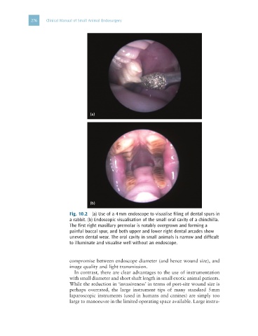
Fig. 10.2 (a) Use of a 4 mm endoscope to visualise filing of dental spurs in
a rabbit. (b) Endoscopic visualisation of the small oral cavity of a chinchilla.
The first right maxillary premolar is notably overgrown and forming a
painful buccal spur, and both upper and lower right dental arcades show
uneven dental wear. The oral cavity in small animals is narrow and difficult
to illuminate and visualise well without an endoscope.
compromise between endoscope diameter (and hence wound size), and
image quality and light transmission.
In contrast, there are clear advantages to the use of instrumentation
with small diameter and short shaft length in small exotic animal patients.
While the reduction in ‘invasiveness’ in terms of port-site wound size is
perhaps overrated, the large instrument tips of many standard 5 mm
laparoscopic instruments (used in humans and canines) are simply too
large to manoeuvre in the limited operating space available. Large instru-